Intensity-Modulated Radiotherapy versus Three-Dimensional Conformal Radiotherapy in Definitive Chemoradiotherapy for Cervical Esophageal Squamous Cell Carcinoma: Comparison of Survival Outcomes and Toxicities
Article information
Abstract
Purpose
The purpose of this study was to compare the survival and toxicities in cervical esophageal squamous cell carcinoma (CESCC) treated by concurrent chemoradiothrapy with either three-dimensional conformal radiotherapy (3D-CRT) or intensity-modulated radiotherapy (IMRT) techniques.
Materials and Methods
A total of 112 consecutive CESCC patients were retrospectively reviewed. 3D-CRT and IMRT groups had been analyzed by propensity score matching method, with sex, age, Karnofsky performance status, induction chemotherapy, and tumor stage well matched. The Kaplan-Meier method and Cox proportional hazards model were used for overall survival (OS) and progression-free survival (PFS). Toxicities were compared between two groups by Fisher exact test.
Results
With a median follow-up time of 34.9 months, the 3-year OS (p=0.927) and PFS (p=0.859) rate was 49.6% and 45.8% in 3D-CRT group, compared with 54.4% and 42.8% in IMRT group. The rates of grade ≥ 3 esophagitis, grade ≥ 2 pneumonitis, esophageal stricture, and hemorrhage were comparable between two groups, while the rate of tracheostomy dependence was much higher in IMRT group than 3D-CRT group (14.3% vs.1.8%, p=0.032). Radiotherapy technique (hazard ratio [HR], 0.09; 95% confidence interval [CI], 0.01 to 0.79) and pretreatment hoarseness (HR, 0.12; 95% CI 0.02 to 0.70) were independently prognostic of tracheostomy dependence.
Conclusion
No survival benefits had been observed while comparing IMRT versus 3D-CRT in CESCC patients. IMRT with fraction dose escalation and pretreatment hoarseness were considered to be associated with a higher risk for tracheostomy dependence. Radiation dose escalation beyond 60 Gy should be taken into account carefully when using IMRT with hypofractionated regimen.
Introduction
Cervical esophageal squamous carcinoma (CESCC) is a relatively rare malignancy, accounting for less than 5% of all esophageal cancers [1]. Historically, surgical resection has been the mainstay of treatment for CESCC, which requires the removal of the larynx, hypopharynx, and esophagus, and deprives these patients of speech and swallowing. As the notion of organ preservation has been introduced and due to the rapid developments in concurrent chemoradiotherapy (CCRT), physicians and patients predisposed to accept the non-aggressive therapy, hoping to improve functional outcomes after therapy. Prior works [2-13] have documented promising overall survival (OS) and functional preservation with definitive CCRT in CESCC patients.
Three-dimensional conformal radiotherapy (3D-CRT) and intensity-modulated radiotherapy (IMRT) are the most effective and commonly used techniques in esophageal cancer. Although recent studies failed to demonstrate the survival benefit of IMRT based definitive CCRT in esophageal cancer [14,15], IMRT does show a greater advantage in target coverage, dose inhomogeneity, and reducing toxicities to normal organs compared to 3D-CRT [16-19]. With the dosimetric advantage of IMRT technique, hypofractionated radiation therapy for various cancers has been comprehensively studied, which shortens the treatment time and intensifies the dose. Several randomized trials demonstrated that dose-escalated, moderately hypofractionated IMRT improves local control in prostate cancer, lung cancer and breast cancer [20-22]. Since previous data suggested that esophageal cancer patients could not benefit from high dose radiation [3], fraction dose escalation (hypofrationated IMRT) started to attract attention especially for CESCC patients.
Since definitive CCRT is an essential treatment option for CESCC, we believed the technique of dose delivery should be well investigated. To our best knowledge, data comparing IMRT with 3D-CRT for CESCC are still limited. Therefore, current study was designed to compare the survival outcomes and toxicities between CESCC patients treated with definitive 3D-CRT and IMRT with concurrent chemotherapy. A propensity score matching (PSM) approach wad used to reduce bias introduced by the non-random treatment assignment, by matching patients with similar clinical stages and other baseline characteristics.
Materials and Methods
1. Patient and clinical data
From January 1, 2000, to September 30, 2016, a total of 138 consecutive patients with CESCC who were treated at the Department of Radiation Oncology, Sun Yat-sen University Cancer Center were retrospectively reviewed. To be included in our study, patients had to meet the following criteria: pathologically confirmed CESSC; treated with definitive radiotherapy with concurrent chemotherapy; without a history of prior radiotherapy; and Karnofsky performance status (KPS) ≥ 70. Surgical therapy was not allowed to be used. Patients with metastasis upper mediastinal lymph nodes (M1 lymph/stage IV) were included. Each patient underwent a physical examination, laboratory tests, electrocardiogram, lung function test, barium contrast study, endoscopy, and computed tomography (CT) scan of the neck, chest, and upper abdomen. Clinical data collected from each patient included age, sex, KPS, smoking or alcohol-abusing history, primary esophageal tumor location, tumor stage, T and N category of primary tumor, total radiation dose, fraction dose, radiotherapy technique, concurrent chemotherapy regimen, pretreatment hoarseness, pretreatment vocal cord paralysis, tracheostomy dependence after treatment, therapeutic toxicities, and tumor progression. Tumor stage was classified using the TNM staging system proposed by the American Joint Committee on Cancer (6th edition).
2. Treatment
All patients received external beam radiation, either 3D-CRT or IMRT, using a 6-8 MV photon beam, with concurrent chemotherapy. During radiotherapy, immobilization, simulation, and treatment planning were performed according to the standard protocol in our department for patients with cervical esophageal carcinoma [23]. The gross tumor volume (GTV) was defined as visible primary tumors (GTV-T) and involved lymph nodes (GTV-N) on endoscopy, CT and/or positron emission tomography (PET) scans. The criteria of lymph node positivity included: short-axis size ≥ 10 mm, or an infiltrative margin, or central necrosis on pretreatment CT scan, reported positive on the pretreatment PET scan, or biopsy positive. The clinical target volume (CTV) included GTV-T with a 3-cm proximal and distal margin, GTV-N and elective nodal regions, including bilateral levels II-IV of the cervical lymph node area and supraclavicular fossa. The planning target volume (PTV) 1 and PTV2 was defined as a 0.5 cm margin added to GTV and CTV, respectively. The radiation doses of 60-70 Gy to PTV1 and 46-54 Gy to PTV2 were delivered in 28-35 fractions, with 5 fractions per week.
Concurrent chemotherapy regimens consisted either of a single agent such as 5-fluorouracil or platinum, or of double agents combined with taxane and platinum (TP) or platinum and 5-fluorouracil (PF).
3. Follow-up and treatment response assessments
The beginning of the follow-up period was defined as the date of diagnosis. All patients were evaluated weekly during therapy, and then underwent a neck and chest or upper abdomen CT scan every 3 months, and an upper digestive tract endoscopy every 6 months for the first 2 years after completion of CCRT, and subsequently every 6 months thereafter until tumor progression. Bone scans were administered when patients were suspected to have bone metastasis. PET scans were performed in patients with suspected systemic progression. The date of last follow-up was January 1, 2018. Treatment response, OS, and progression-free survival (PFS) were recorded. The responses to CCRT were assessed 2 months after radiotherapy by an independent radiation oncologist according to Response Evaluation Criteria in Solid Tumors ver. 1.1. OS was calculated from the diagnosis of CESCC until death or last follow-up. PFS was defined as the time from diagnosis to tumor recurrence, or death, or last follow-up. OS and PFS were used as measures of survival outcomes. Acute and late toxicities were collected retrospectively and then presented according to The National Cancer Institute Common Toxicity Criteria (ver. 4.0).
4. Statistical analysis
We matched patients in the 3D-CRT and IMRT groups by propensity score with a caliper of 0.1 in a 1:1 ratio, using the PSM method with five covariates including sex, age, KPS, induction chemotherapy, and tumor stage. Means, frequencies, and percentage were calculated to describe the data. Continuous variables, such as age, total radiation dose, fraction dose, D5 of PTV1, GTV volume, Dmean of larynx and so on, were normalized as the sample median and then analyzed as nominal categorical variables. Comparisons of categorical variables between groups were first performed by Fisher exact test, and then the variables that reached a p-value < 0.1 were further evaluated in a multivariate analysis using Logistic regression model. The Kaplan-Meier method was used to produce survival curves. The 3-year, 5-year OS and PFS were estimated. OS and PFS were first compared in univariate analysis by using the two-sided logrank test. Then the Cox proportional hazards model was used to test independent prognostic factors of OS and PFS. All statistical analyses were performed using SPSS ver. 24.0 software (IBM Corp., Armonk, NY), and differences were considered significant at a p-value < 0.05.
5. Ethical statement
Our study was approved by the Ethics Committee of Sun Yat-sen University Cancer Center, and the approval number was YB2017-080. As this was a retrospective analysis of routine clinical data, a waiver of the requirement for individual informed consent was granted by our institutional ethics committee.
Results
1. Baseline characteristics
A total of 112 of the included 138 consecutive patients were divided into the 3D-CRT and IMRT groups based on the above-mentioned PSM procedure. Patient sex, age, KPS, induction chemotherapy, and tumor stage were well matched (p > 0.1). The demographic and clinical characteristics of the cohort are listed in Table 1. The median age was 59 years, range from 18 to 81. Most patients (102/112, 77%) had stages II and III disease and the rest 10 patients had stages IV disease. As for T category, 21 patients had T1-2 lesion, and 89 had T3-4 lesion. In our group, 9.8% (11/112) patients had pretreatment hoarseness, and the incidence of pretreatment vocal cord paralysis was 6.3% (7/112). The median radiation dose was 62 Gy (range, 60 to 70 Gy) in 30-35 fractions. The dosimetric parameters were listed in Table 2. The median fraction dose, Dmean of GTV, V5 of the lungs, V20 of the lungs, Dmean of the lungs, Dmax of the spinal cord, D5 of PTV1 and Dmean of larynx in the 3D-CRT group vs. IMRT group was 200 cGy vs. 213 cGy (p < 0.001, Fisher exact test), 6,123 cGy vs. 6,290 cGy (p=0.001), 38.0 vs. 50.2 (p=0.032), 18.7 vs. 21.8 (p=0.479), 958 cGy vs. 1,101 cGy (p=0.156), 4,917 cGy vs. 4,344 cGy (p < 0.001), 6,394 cGy vs. 6,745 cGy (p=0.007), and 5,779 cGy vs. 6,045 cGy (p=0.227), respectively. Most patients (93/112, 83%) received double agents chemotherapy regimens, and the remaining adopted single-agent regimens. Part of patients (27/112, 24%) underwent induction chemotherapy before definitive CCRT.
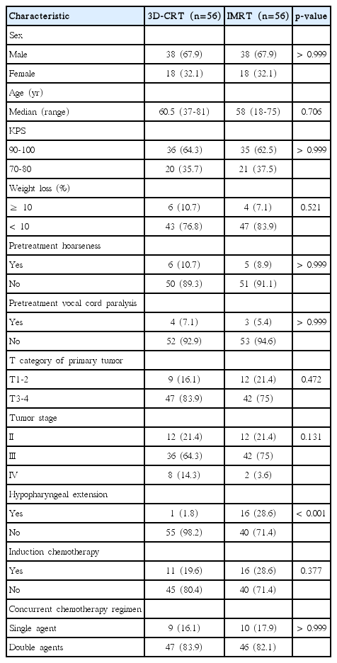
Demographic and clinical characteristics of the matched patients
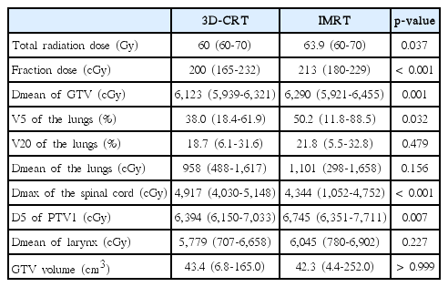
Dosimetric parameters
2. Survival outcomes
With a median follow-up of 34.9 months (range, 2.1 to 183.6 months), our analysis demonstrated a median estimated OS of 36.0 months in the 3D-CRT group, 45.6 months in the IMRT group and 41.4 months in the whole group. The 3-year and 5-year OS rate was 49.6% and 45.6% in the 3D-CRT group, compared with 54.4% and 43.8% in the IMRT group (p=0.927, log-rank). The median estimated PFS was 30.2, 25.2, and 27.5 months in the 3D-CRT, IMRT, and the whole group, respectively. And the 3-year and 5-year PFS rate was 45.8% and 32.1% in the 3D-CRT group, vs. 42.8% and 32.1% in the IMRT group (p=0.859, log-rank) (Fig. 1). There was no statistically significant difference observed in OS and PFS between the 3D-CRT group and the IMRT group.

Overall survival (OS) (A) and progression-free survival (PFS) (B) between the three-dimensional conformal radiotherapy (3D-CRT) and intensity-modulated radiotherapy (IMRT) groups. There was no statistically significant difference observed in OS (p=0.927, log-rank) and PFS (p=0.859, log-rank) between the 3D-CRT group and the IMRT group.
3. Prognostic factors
Univariate Kaplan-Meier survival analysis found that sex (p=0.088) and tumor stage (p=0.053) were significantly associated with OS (Table 3). Multivariate Cox proportional hazards model identified none of them reached statistical significance (Table 4). Similarly, univariate analysis of factors influencing PFS showed that sex (p=0.058), concurrent chemotherapy regimen (p=0.034), induction chemotherapy (p=0.054) and tumor stage (p < 0.001) were statistically significant variables. Only sex remained independently related to PFS in multivariable analysis (hazard ratio [HR], 0.60; 95% confidence interval [CI], 0.36 to 0.99).
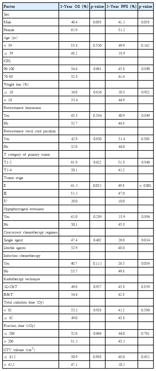
Univariate analysis of prognostic factors for OS and PFS

Multivariate analysis of prognostic factors for OS and PFS
4. Toxicities
The most commonly documented therapeutic toxicity was radiation esophagitis, and most patients (60/112, 53.6%) had grade 3 esophagitis. Seventeen percent of patients (19/112) had esophageal stricture and required dilatation, with 12 in the 3-dimensional radiotherapy (3D-RT) group and seven in the IMRT group. Other observed toxicities were mostly grade 1 or 2, including radiation pneumonitis and gastrointestinal toxicity. There were both one reported case of life-threatening hemorrhage in the 3D-CRT group and the IMRT group. However, none of these toxicities reached statistical significance between the 3D-RT group and the IMRT group (Table 5). No patient developed acute grade 4 toxicity, and there were no treatment-related deaths.
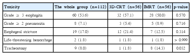
Univariate analysis of therapeutic toxicities between 3D-CRT and IMRT
Moreover, we observed that 8.0% of patients (9/112) developed dyspnea and required tracheostomy within 6 months after cessation of therapy, without any evidence of tumor progression or second primary tumor. Univariate analysis indicated that radiotherapy (RT) technique (p=0.032), fraction dose (p=0.091), pretreatment hoarseness (p=0.043), and pretreatment vocal cord paralysis (p=0.098) were significantly related to tracheostomy dependence (Table 6). IMRT, fraction dose more than 206 cGy, pretreatment hoarseness, and pretreatment vocal cord paralysis were risk factors for tracheostomy dependence. Since fraction dose was significantly associated with RT technique (p < 0.001), and pretreatment vocal cord paralysis was significantly associated with pretreatment hoarseness (p < 0.001), we excluded fraction dose and pretreatment vocal cord paralysis from multivariate analysis, then the other two variables reached p < 0.1 in univariate analysis were further analyzed by using a Logistic regression model. RT technique (HR, 0.09; 95% CI, 0.01 to 0.79) and pretreatment hoarseness (HR, 0.12; 95% CI, 0.02 to 0.70) were independently prognostic of tracheostomy dependence (Table 7).

Univariate analysis of prognostic factors for tracheostomy dependence
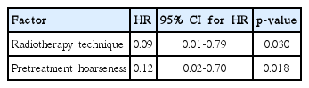
Multivariate analysis of prognostic factors for tracheostomy dependence
Discussion
Although dosimetric studies [16-19] reported that IMRT was better than 3D-CRT with respect to improved target coverage and conformality, the clinical benefits of IMRT have not been well established. To our knowledge, this is the first study to compare the survival outcomes and toxicities of CESCC between the 3D-CRT and the IMRT group using PSM approach with the largest sample size. Our study indicated OS and PFS were comparable between the 3D-CRT and IMRT groups. The 3-year and 5-year OS rate was 49.6% and 45.6% in the 3D-CRT group, while 54.4% and 43.8% in the IMRT group (p=0.927, log-rank). The 3-year and 5-year PFS rate was 45.8% and 32.1% in the 3D-CRT group, vs. 42.8% and 32.1% in the IMRT group (p=0.859, log-rank).
Advances in radiotherapy technique in treatment planning and radiation delivery, such as simultaneous integrated boosting IMRT, could allow the optimization of target volume coverage, which theoretically resulted in better survival outcomes. However, the published data failed to support the significant advantage of IMRT in survival. Yang et al. [24] reported that OS and failure-free survival (FFS) were not significantly different between the IMRT and 3D-CRT group, with the 2-year OS and FFS for the 3D-CRT vs. IMRT group was 53.6% vs. 55.6% (p=0.965), and 49.5% vs. 56.7% (p=0.998), respectively. Similarly, there was no statistically significant difference observed in OS, local FFS, and regional FFS between the IMRT group and two-dimensional conformal radiotherapy group in the study conducted by Cao et al. [25]. A recent published data from Japan [26] showed that IMRT achieved a significantly better 3-year OS than 3D-CRT (81.6% vs. 57.2%, p=0.037). They found that the sufficient salvage rate was higher in IMRT than 3D-CRT group when locoregional recurrence was presented, which contributed to the difference in OS. There was yet no statistical difference in locoregional control or PFS between these two groups. Similarly to most of the previous studies, our study did not support the survival advantage of IMRT compared with 3D-CRT. In our study, the Dmean of GTV was higher in IMRT group (6,290 cGy vs. 6,123 cGy, p=0.001). It suggests that there was no more dose-response relationship in dose ranges over 50 Gy, as indicated by series of Radiation Therapy Oncology Group studies [2,3].
IMRT could achieve well-demonstrated sparing of organs at risk, such as lungs and spinal cord, reducing both acute and late toxicities. However, our analysis showed that the rates of observed toxicities, including pneumonitis, esophagitis, esophageal stricture, gastrointestinal reaction, and life-threatening hemorrhage were comparable between the 3D-CRT group and the IMRT group. Detailed dosimetric study showed similar V20 and mean dose of the lungs between two groups, a relatively lower Dmax of spinal cord and a higher V5 of the lungs in the IMRT group. These results indicated that both 3D-CRT and IMRT could be delivered safely without obvious toxicities when the total dose did not exceed 70 Gy.
Within 6 months after CCRT, nine patients (8%, 9/112) developed dyspnea and required tracheostomy. All of the nine patients got partial remission after therapy and did not show any evidence of tumor progression at the time of tracheostomy. Few published reports had recorded and analyzed this complication. Gkika et al. [13] reported one patient (1.8%, 1/55) with CESCC underwent tracheostomy during definitive CCRT because of inability to breathe, who had got a paralysis of the recurrent laryngeal nerve before treatment. In Tong’s study [4], two patients (9.5%, 2/21) treated with up-front CCRT had bilateral vocal cord palsy requiring permanent tracheostomy. Staton et al. [27] reviewed 45 patients with advanced laryngeal cancer at 6 months after CCRT, and they found that the rate of tracheostomy dependence was 44% in patients with baseline vocal fold fixation versus 6% in patients without fixation. The authors attributed tracheostomy dependence to radiation-related bilateral recurrent laryngeal nerve paralysis, which results in bilateral vocal cord fixation. Similarly, in our cohort, pretreatment hoarseness was an independent risk factor for tracheostomy dependence during or after CCRT. As a result, it is urgent to call for close surveillance in those with pretreatment hoarseness or vocal cord paralysis during and 6 months after therapy. Our results showed a significantly higher rate of tracheostomy in IMRT group than that in 3D-CRT group (14.3% vs. 1.8%, p=0.032). Most IMRT plans in our cohort utilize the SIB technique with the median fraction dose to GTV of 213 cGy. For patients with CESSC, recurrent laryngeal nerves are always located in the proximity of GTV, which inevitably received a fraction dose more than 200 cGy when treated with IMRT. Therefore, we inferred that IMRT with fraction dose escalation (hypofractionated IMRT) might lead to a higher rate for tracheostomy dependence. Hypofractionated IMRT scheme should be carefully defined especially with concurrent chemotherapy in case of severe adverse events.
This retrospective study has several limitations, such as selection bias and relatively small number of patients which might affect the results of our study, and it was a single-institution experience. Larger multi-center prospective trials are warranted to validate our results.
No survival benefits had been observed while comparing IMRT versus 3D-CRT in CESCC patients. Sex was the only independent prognostic factor for PFS. IMRT with fraction dose escalation (hypofractionated IMRT) was considered to be associated with a higher risk for tracheostomy dependence within 6 months after definitive CCRT, and patients with pretreatment hoarseness or vocal cord paralysis should be under close surveillance for dyspnea. Radiation dose escalation beyond 60 Gy should be taken into account carefully when using IMRT with hypofractionated regimen. Prospective studies are warranted to verify presented results.
Notes
Conflict of interest relevant to this article was not reported.
Acknowledgements
Our study was supported by Guangdong Esophageal Cancer Institute Science and Technology Program (M201505).