Prognostic Significance of Retroperitoneal Lymphadenectomy, Preoperative Neutrophil Lymphocyte Ratio and Platelet Lymphocyte Ratio in Primary Fallopian Tube Carcinoma: A Multicenter Study
Article information
Abstract
Purpose
The purpose of this study is to evaluate the prognostic role of preoperative neutrophil to lymphocyte ratio (NLR) and platelet to lymphocyte ratio (PLR) and the need for para-aortic lymphadectomy in patients with primary fallopian tube carcinoma (PFTC).
Materials and Methods
Ninety-one patients with a diagnosis of PFTC were identified through the gynecologic oncology service database of six academic centers. Clinicopathological, surgical, and complete blood count data were collected.
Results
In univariate analysis, advanced stage, suboptimal surgery, and NLR > 2.7 were significant prognostic factors for progression-free survival, whereas in multivariate analysis, only advanced stage and suboptimal surgery were significant. In addition, in univariate analysis, cancer antigen 125 ≥ 35 U/mL, ascites, advanced stage, suboptimal surgery, NLR > 2.7, PLR > 233.3, platelet count ≥ 400,000 cells/mm3, staging type, and histological subtype were significant prognostic factors for overall survival (OS); however, in multivariate analysis, only advanced stage, suboptimal surgery, NLR > 2.7, and staging type were significant. Inclusion of pelvic and para-aortic lymphadenectomy in surgery showed significant association with longer OS, with a mean and median OS of 42.0 months and 35.5 months (range, 22 to 78 months), respectively, vs. 33.5 months and 27.5 months (range, 14 to 76 months), respectively, for patients who underwent surgery without para-aortic lymphadenectomy (hazard ratio, 3.1; 95% confidence interval, 1.4 to 5.7; p=0.002).
Conclusion
NLR (in both univariate and multivariate analysis) and PLR (only in univariate analysis) were prognostic factors in PFTC. NLR and PLR are inexpensive and easy tests to perform. In addition, patients with PFTC who underwent bilateral pelvic and para-aortic lymphadenectomy had longer OS.
Introduction
Primary fallopian tube carcinoma (PFTC) is a rare neoplasm, accounting for 0.14%-1.8% of all gynecological malignancies [1,2]. However, recent data suggest that the true incidence of PFTC has been substantially underestimated; this conclusion is based on compelling evidence that papillary serous carcinoma, the most common subtype of epithelial ovarian carcinoma (EOC), actually arises from the epithelial lining of the fallopian tube [1]. The most common histological type of gynecological malignancy is serous adenocarcinoma [1]. The mean age at diagnosis of PFTC is 64 years, and incidence peaks at 70-74 years [1,2].
Total abdominal hysterectomy with bilateral salpingooophorectomy and infracolic omentectomy, appendectomy, peritoneal washing, and peritoneal biopsy constitute the primary treatment of choice for PFTC; inclusion of pelvic and para-aortic lymphadenectomy has been controversial [3]. Klein et al. [4] reported that routine retroperitoneal sampling and node dissection are necessary for proper staging, whereas other authors have reported that systemic retroperitoneal lymphadenectomy is preferable due to the strong tendency of the tumor to spread in the lymphatic system [5]. In addition, the therapeutic value of a systematic complete lymphadenectomy remains controversial, with a paucity of clinical data to support its use [2].
The overall 5-year survival for patients with PFTC is 22%-57% [6]. Many prognostic factors have been investigated in an effort to better estimate patient outcome. Stage, patient age, and residual tumor after initial surgery are consistently important prognostic factors. In addition, according to some reports, a closed fimbriated end of the fallopian tube, positive peritoneal cytology, lesion site within the tube (fimbrial vs. non-fimbrial), human epidermal growth factor receptor 2/nu-positive expression, p53 alteration, elevated pretreatment cancer antigen 125 (CA-125), and lymphovascular space involvement are also important prognostic factors [2-6]. Interest in elevated neutrophil to lymphocyte ratio (NLR) and platelet to lymphocyte ratio (PLR) as markers of systemic inflammatory response (SIR) in various clinical circumstances is increasing. In recent clinical studies, these ratios have been investigated as advanced-stage predictors or prognostic factors in different types of gynecologic cancer [7-14]. In addition, preoperative thrombocytosis has been associated with adverse outcomes from endometrial and ovarian cancers [15,16]. However, no study investigating the predictive value of NLR, PLR, or thrombocytosis in overall survival (OS) and disease-free survival of patients with PFTC has been reported.
Therefore, we retrospectively reviewed the clinical characteristics, management, and outcomes of 91 patients with PFTC who were treated in six gynecological oncology departments. In addition, this study sought to determine whether 1) NLR, PLR, or thrombocytosis plays a role in disease-free survival and OS of PFTC and 2) performance of para-aortic lymphadenectomy is essential for patients with PFTC.
Materials and Methods
The databases of six gynecological oncology departments from Turkey, including Izmir Tepecik Education and Research Hospital, Izmir Ege University School of Medicine, Eskisehir Osmangazi University School of Medicine, Ankara University School of Medicine, Antalya Akdeniz University School of Medicine, and Adana Cukurova University School of Medicine were reviewed for identification of patients with complete information and pathologically proven PFTC who underwent surgical staging between January 1, 1996, and December 31, 2011. This study was conducted in accordance with the ethical standards of the Declaration of Helsinki and was approved by the local ethics committees of Izmir Tepecik Education and Research Hospital.
Cases were identified according to the PFTC diagnostic criteria established by Hu et al. [17] and modified by Sedlis [18]. All slides were reviewed by expert pathologists from each institution. The following clinical data were collected from patient medical, surgical, pathological, and chemotherapy reports: demographic characteristics, presenting symptoms, serum CA-125 level, preoperative complete blood cell count (CBC), date and type of surgical procedure, presence or absence of residual tumor after surgery, number of excised and positive lymph nodes, presence or absence of ascites, tumor pathological characteristics (grade and size), type of first-line chemotherapy, date of recurrence, treatment after recurrence, date of last medical examination, and date of death.
The exclusion criteria were neoadjuvant chemotherapy and tumors of low malignant potential. Patients with any inflammatory signs or conditions, hematological disease, or other malignancies that might affect the CBC results, smokers, those who used corticosteroids or β-agonists, and patients with missing data including preoperative CBC and surgico-pathological notes, were excluded. International Federation of Gynecology and Obstetrics (FIGO) staging according to the 2009 revised classification system was used. Patients with stage IC-IV disease received chemotherapy following surgery.
CBC was measured within one month preoperatively using a Coulter LH 750 device (Beckman Coulter, Brea, CA) or a Sysmex XE-2100 device (TOA Medical Electronics, Kobe, Japan) at the six centers. When more than one CBC result was available, the result from the date closest to staging surgery was recorded for statistical analysis. The NLR was defined as the absolute neutrophil count divided by the absolute lymphocyte count, and the PLR was defined as the absolute platelet count divided by the lymphocyte count.
Patients were classified as not having been staged if only a unilateral salpingo-oophorectomy, total hysterectomy with unilateral or bilateral salpingo-oophorectomy with or without omentectomy was performed. Partial staging was defined as pelvic washing, peritoneal biopsy, omentectomy, bilateral pelvic lymph node dissection with a unilateral salpingo-oophorectomy, or total hysterectomy with unilateral or bilateral salpingo-oophorectomy. Complete staging was defined as pelvic washing, peritoneal biopsy, omentectomy, bilateral pelvic and para-aortic lymph node dissection with a unilateral salpingo-oophorectomy, or total abdominal hysterectomy with unilateral or bilateral salpingo-oophorectomy.
Optimal debulking was defined as a procedure that left a maximum residual tumor < 1 cm in diameter. Thrombocytosis was defined as a platelet count ≥ 400,000 cells/mm3. Patients returned for a follow-up evaluation every three months for the first 2 years, every 6 months for the next 3 years, and annually thereafter. Computed tomography or magnetic resonance imaging was performed annually. Survival data were analyzed in December 2011. The survival analysis was based on the Kaplan-Meier method, and the results were compared using the log-rank test. Progressionfree survival (PFS) was defined as the time from the date of primary surgery to detection of recurrence or the latest observation. OS was defined as the time from the date of primary surgery to death or the latest observation. The chi-square test and Student’s t test for unpaired data were used for the statistical analysis. Cox regression analysis was used to determine factors affecting survival, and results are presented as hazard ratios (HRs) with 95% confidence intervals (CIs). All statistical analyses were performed using MedCalc software ver. 11.5 for Windows (MedCalc, Mariakerke, Belgium). A p-value of < 0.05 was considered significant.
Results
Ninety-one women with PFTC were identified during the study period. The characteristics of the patients are shown in Table 1. The mean age was 56.9 years (range, 32 to 81 years). None of the patients was correctly diagnosed with PFTC preoperatively. The two most common preoperative diagnoses were ovarian carcinoma (42.9%) and adnexal mass of unknown nature (29.7%). The histopathological characteristics of the patients are shown in Table 2. Papillary serous histology (67%) was the predominant histological subtype.
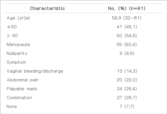
Clinical characteristics of the study population
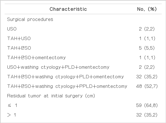
Surgical and clinicopathologic characteristics of the patients
The median follow-up period was 34 months (range, 14 to 78 months). Thirty-four patients (37.4%) had FIGO stage I disease, 10 (10.9%) had stage II, 43 (47.3%) had stage III, and four (4.4%) had stage IV disease. Nine patients (9.9%) had no surgical staging, 34 (37.4%) had partial staging, and 48 (52.7%) had complete staging. A detailed description of the operative procedures performed is shown in Table 2. Optimal surgery with residual tumor ≤ 1 cm was achieved in 64.8% of cases.
Seventy-nine patients (86.8%) received adjuvant chemotherapy after primary surgery. Patients receiving adjuvant therapy had stage IC or higher disease. None of the patients had undergone a secondary staging procedure following primary surgery. Four unstaged patients who refused reoperation were treated based on high-risk features such as grade 3 disease or tumor rupture.
We studied NLR and PLR at different levels to determine the optimal cut-off point for prediction of disease stage (early vs. advanced) and primary surgical outcome (optimal vs. suboptimal). The receiver operating characteristics (ROC) curve indicated that NLR > 2.7 and PLR > 233.3 was optimum for prediction of advanced stage (stage III/IV) and suboptimal surgery (Table 3).

Predictive values of NLR and PLR for determination of disease stage and optimality of surgery
The mean and median PFS were 34.2 months and 32 months (range, 9 to 76 months), respectively. Results of univariate and multivariate analyses for PFS are shown in Table 4. Results of univariate analysis indicated that advanced FIGO stages III and IV, suboptimal surgery, and pretreatment NLR > 2.7 (Fig. 1A and C) were adverse prognostic factors for PFS. Results of multivariate analysis showed that advanced FIGO stage and suboptimal surgery were independent prognostic factors for PFS.
Results of univariate and multivariate analyses of disease-free survival of patients with fallopian tube carcinoma
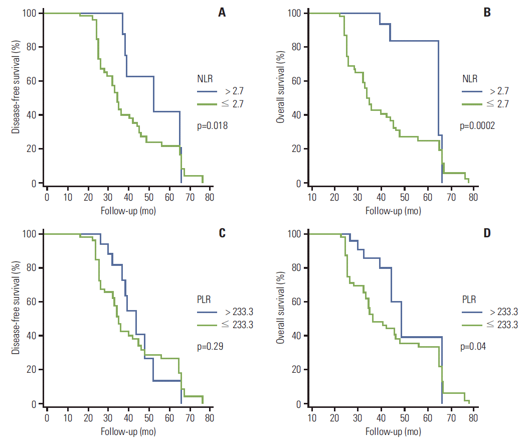
(A) Disease-free survival curves according to neutrophil to lymphocyte ratio (NLR). (B) Overall survival curves according to NLR. (C) Disease-free survival curves according to platelet to lymphocyte ratio (PLR). (D) Overall survival curves according to PLR.
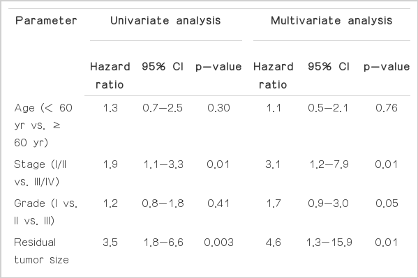
The 5-year OS rate was 63.7%, and the mean and median survival durations were 38.9 months and 35 months (range, 14 to 78 months), respectively. Results of univariate and multivariate analyses for OS are shown in Table 5. In univariate analysis of survival rates, pretreatment CA-125 ≥ 35 U/mL, presence of ascites, advanced FIGO stage, suboptimal surgery, pretreatment NLR > 2.7 (Fig. 1B), pretreatment PLR >233.3 (Fig. 1D), pretreatment platelet count ≥ 400,000 cells/mm3, surgical staging type (none vs. partial vs. complete), and histological subtype (serous vs. non-serous) showed significant association with poor OS. In contrast, multivariate analysis of OS showed significant association of advanced FIGO stage, suboptimal surgery, pretreatment NLR > 2.7, and surgical staging type (none vs. partial vs. complete) with poor OS.

Results of univariate and multivariate analyses for overall survival of patients with fallopian tube carcinoma
Of all patients, 82 (90.1%) underwent surgical staging procedures. Of these, 48 (58.6%) had complete staging, and 34 (41.4%) had incomplete staging. The mean and median PFS tended to be better in the complete staging group (38.1 months and 34.0 months [range, 9 to 76 months], respectively) than in the partial staging group (27.8 months and 24.0 months [range, 11 to 76 months]), however, the difference was not significant (HR, 1.7; 95% CI, 0.9 to 3.2; p=0.09) (Fig. 2A). Complete staging showed significant association with longer OS, with mean and median OS rates of 42.0 months and 35.5 months (range, 22 to 78 months), respectively, vs. 33.5 months and 27.5 months (range, 14 to 76 months), respectively, in the partial staging group (HR, 3.1; 95% CI, 1.4 to 5.7; p=0.002) (Fig. 2B).
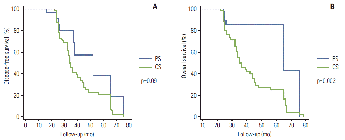
(A) Disease-free survival rates according to surgical staging type (partial staging [PS] vs. complete staging [CS]). (B) Overall survival rates according to surgical staging type (PS vs. CS).
Discussion
The current study retrospectively analyzed patients with PFTC who were treated at six gynecologic oncology centers in Turkey. Our aim was to describe the demographic and clinical characteristics of PFTC and to identify variables affecting PFS and OS in patients with this disease.
PFTC is the least common gynecologic malignancy. Unlike ovarian cancer, it is not routinely suspected in women with a pelvic mass. In our study, none of the patients had a definitive preoperative diagnosis of PFTC. In the literature, the mean age of patients with PFTC is 55 years [6], similar to that in our patient population. Schneider et al. [19] reported that PFTC is diagnosed at an earlier stage than ovarian cancer, possibly as a result of abdominal pain resulting from tubal distention and intermittent serosanguineous discharge.
The 5-year OS rate for all stages of PFTC varies from 22% to 81.7% [20-22]. Our rate was 63.7%. Several studies have reported various prognostic factors for predicting the outcome of PFTC, including age, stage, cell type, elevated CA-125, and residual tumor after surgery [2,5,6]. Shamshirsaz et al. [20] reported that in a univariate analysis, pretreatment serum CA-125 values were prognostic with respect to both PFS and OS. They also showed that the side where the tumor was located was associated with PFS [20]. Alvarado-Cabrero et al. [23] reported significant correlation of tumor grade with survival. However, multivariate analyses were not performed in these two studies [20,23]. In the current study, advanced FIGO stage, presence of a residual tumor > 1 cm after surgery, and surgical staging type (none vs. partial vs. complete) showed significant association with poor OS in both univariate and multivariate analyses. Location and grade of tumor did not show association with PFS or OS in our univariate or multivariate analyses.
Prognostic factors other than age and elevated CA-125 can only be assessed during or after surgery based on the pathological features of the cancer. Do NLR, PLR, and thrombocytosis play a role in disease-free and OS in patients with PFTC? Leukocyte migration is the cornerstone event in the immune response to the neoplastic process [24]. An increase in neutrophil count, a slight increase in platelet count, and a decline in lymphocytes are systemic alterations that are part of the inflammatory response to the growth, progression, and spread of tumors [7,24]. In this respect, interest in NLR and PLR has shown a recent increase, and they have been studied as SIR markers in non-gynecologic and gynecologic cancers, with results suggesting that a preoperative increase in NLR and/or PLR is significantly associated with nodal metastasis and advanced stage and is a significant prognostic factor [8-14]. Indeed, it has been suggested that these ratios, which can be assessed using noninvasive, simple, and inexpensive methods and can be easily calculated from the CBC, should be included in the routine assessment of all patients with cancer [8]. More recently, thrombocytosis was reported as an independent predictor of increased risk of recurrence in 587 women who underwent staging surgery for EOC [15].
Fallopian tube carcinomas have a similar histology to their ovarian counterparts. To date, only four studies have analyzed pretreatment NLR and/or PLR values in patients with EOC [11-14], and the value of these ratios as prognostic factors in patients with PFTC has not been validated. A preoperative NLR cut-off point > 2.6 distinguished ovarian cancer from benign conditions and predicted adverse outcomes [11], and a similar ratio was associated with advanced-stage disease or suboptimal ovarian surgery [12]. PLR was superior to NLR for prediction of poor survival in two ovarian cancer studies [13,14]. First, Asher et al. [13] showed that PLR > 300 was superior to NLR > 4.0 for prediction of poor survival and was a novel independent prognostic marker for EOC. Later, it was demonstrated that PLR >200 had the potential to predict advanced-stage disease or suboptimal surgery and was a better prognostic indicator for survival of patients with EOC than NLR of 2.6 [14].
In our study, results of the ROC curve analysis showed that the optimum cut-off value for prediction of advanced stage (stage III/IV) and suboptimal surgery in patients with PFTC was 2.7 for NLR, with 85.1% sensitivity and 75.0% specificity for advanced stage; suboptimal surgery and a PLR value of 233.3 showed 77.9% sensitivity and 84.3% specificity, with 87.2% sensitivity and 63.4% specificity for advanced stage and 81.3% sensitivity and 71.8% specificity for suboptimal surgery. In a univariate Cox survival analysis, these ratios and a pretreatment platelet count ≥ 400,000 cells/mm3 showed significant association with poor OS. However, in the multivariate Cox survival analysis, NLR > 2.7 was also a significant prognostic marker, along with stage and postoperative residual disease. Our results are consistent with those of previous studies and support the growing notion that the inflammatory process is linked to adverse outcomes in pelvic serous tumors.
Is performance of a para-aortic lymphadenectomy essential for PFTC? Due to its rarity, optimal management for patients with PFTC has not been well defined, and there is a tendency to treat this malignancy like ovarian cancer [21]. The need for complete surgical staging, including lymph node sampling or dissection, is controversial [3,22,25]. These procedures may be performed in different ways by different surgeons in many centers; some highlight early extensive lymphadenectomy, some perform only pelvic lymphadenectomy, and others favor lymph node sampling or do not always perform lymphadenectomy [2,3]. In our study, in univariate and multivariate analyses, type of staging surgery (no staging, partial, or complete) showed significant association with OS.
Koo et al. [3] reported that para-aortic lymphadenectomy is an important procedure in patients with PFTC and suggested that para-aortic nodes are the sentinel lymph nodes in these patients. In our analysis of patients treated with pelvic lymphadenectomy with or without para-aortic lymphadenectomy (partial vs. complete staging), we found that the complete staging group showed significantly longer OS compared with the partial staging group. The mean and median PFS tended to be better in the complete staging group than in the partial staging group, but the difference was not significant.
Potential limitations of this study include its retrospective nature, the use of two different devices for blood sample analysis for calculation of NLR, PLR, and platelet counts, and the evaluation of data without preoperative C-reactive protein results because this test is not performed routinely at the six centers. Despite these limitations, the availability of good follow-up data and one of the largest PFTC cases series should increase the validity of results and mitigate the weaknesses.
Conclusion
The following conclusions can be drawn from our data. First, in univariate analysis, only pretreatment NLR > 2.7 was found to be an adverse prognostic factor for PFS. However, in univariate analysis, pretreatment NLR > 2.7, pretreatment PLR > 233.3, and pretreatment platelet count ≥ 400,000 cells/mm3 showed significant association with poor OS. In addition, pretreatment NLR > 2.7 was an independent prognostic factor for OS. NLR and PLR are inexpensive and easy tests to perform, and they provide useful prognostic information. Finally, patients who underwent complete staging surgery for PFTC had longer PFS and OS.
Notes
Conflict of interest relevant to this article was not reported.