Pleural Metastasis as Initial Presentation of Occult Gastric Cardia Cancer: A Possible Role of PET-CT in Diagnosis
Article information
Abstract
We report on a case of malignant pleural effusion as initial metastatic presentation of occult gastric cardia cancer in a young woman. To the best of our knowledge, this is the first report of gastric adenocarcinoma metastasized to pleura as an initial presentation. Location of cardia and signet ring cell histology may contribute to the manifestation. Utilization of positron emission tomography-computed tomography was helpful for proper diagnosis. For patients with such distinct clinical presentations, it would be appropriate to consider gastric cancer as one of the possible primary sites.
Introduction
Gastric cancer is one of the most common malignancies in Eastern Asia. In advanced stage cancer, common metastatic sites include distant lymph nodes, liver, and peritoneum, with rare metastasis to lung, bone, brain, and leptomeningeal space. We report here on an unusual clinical presentation of occult gastric cancer in a relatively young woman.
Case Report
A 44-year-old woman visited our center for dyspnea in June 2012. A large amount of both side pleural effusions and moderate pericardial effusions were detected on chest radiography and computed tomography (CT). Pleural fluid analysis showed exudative type effusion, however, the etiology was not clear. There was no definite evidence of tuberculous pleurisy. Repeated cytologic examination was also non-diagnostic.
Pleural effusion persisted despite repeated drainage and empirical anti-tuberculosis medications were not effective. We performed pleural biopsy with closed tube thoracotomy at the right side pleural cavity, which revealed a signet ring cell carcinoma (Fig. 1A). We performed a series of work-ups to find the primary site.

(A) Pleural biopsy shows clusters of poorly differentiated carcinoma with signet ring cell features, infiltrating into the skeletal muscle fibers (H&E staining, ×200). (B) The biopsy shows clusters of poorly differentiated carcinoma with signet ring cell features and remaining gastric foveolar epithelium (upper part) (H&E staining, ×200).
Chest CT scan showed no abnormal findings except bilateral pleural and pericardial effusion and there were no lesions suggesting malignancy in both lung parenchyma. Abdomen CT scan also showed no abnormal lesion but only a benign looking ovarian cyst.
Gastrofiberscopy showed no particular findings except atrophic gastritis at the antral area.
With the possibility of primary lung signet ring cell carcinoma, although there was no lesion in the lung, we checked ALK translocation by fluorescence in situ hybridization in the biopsy tissue, which was negative [1].
Serum tumor marker study was not helpful either. Carcinoembryonic antigen and cancer antigen (CA) 15-3 levels were both normal and CA 125 and CA 19-9 levels were only minimally increased (36.38 U/mL and 55.52 U/mL, respectively).
Positron emission tomography-computed tomography (PET-CT) showed a hypermetabolic lesion at the right pleural area at the sixth rib (maximum standardized uptake value [SUVmax], 6.1), gastric cardia (Fig. 2A), and right ovary, the latter two of which were regarded as an inflammatory or non-specific uptake at that time.
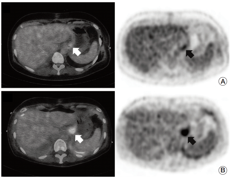
(A) Mild fludeoxyglucose uptake (maximum standardized uptake value [SUVmax], 4.3) seen at gastric cardia. (B) Larger and more prominent uptake (SUVmax, 8.0) seen four months later.
With the diagnosis of carcinoma of unknown primary, we administered paclitaxel plus carboplatin combination chemotherapy. After two cycles, there was no improvement of clinical symptoms and pleural effusion persisted. We switched the chemo-regimen to pemetrexed plus cisplatin and treated her with another three cycles and performed repeat PET-CT for evaluation of disease status. With aggravated pleural effusion, the hypermetabolic lesion at the cardia became prominent (SUVmax, 8.0) (Fig. 2B). Fludeoxyglucose uptake of the effusions including a previous right pleural lesion around the sixth rib also increased. With gastrofiberscopic biopsy, we could confirm cardia cancer pathologically. It was signet ring cell carcinoma, the same histologic type (Fig. 1B). Even at that time, the endoscopic finding was not so revealing. There was only a hyperemic erosive lesion in the cardia (Fig. 3). Careful review of the photos recorded from the first gastrofiberscopy found no significant lesions in the cardia.
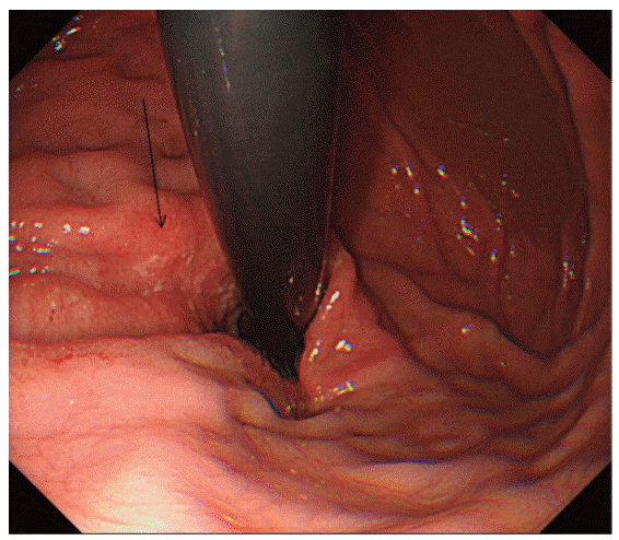
Endoscopic finding of a patient with ill-defined hyperemic erosions in the cardia at retroflexion view.
We treated the patient with 5-fluorouracil/leucovorin plus oxaliplatin (FOLFOX) combination as a standard chemotherapy protocol of metastatic gastric cancer in our center. Her clinical symptoms showed moderate improvement with this regimen, but only briefly. She eventually expired from disease progression three months after start of FOLFOX chemotherapy in January 2013.
Discussion
Pleural metastasis and malignant pleural effusion occurs with a variety of cancers. Lung cancer, which is responsible for approximately 30% of cases, followed by cancer of the breast, ovaries, and lymphoma, remain the most common causes [2].
In this report, we discovered signet ring cell carcinoma on the pleural biopsy, which originated from occult cardia cancer. To the best of our knowledge, this is the first report of a case of gastric carcinoma metastasized to pleura as an initial dominant presentation.
Gastric cardia cancer is relatively uncommon and rather distinct in its characteristics. According to previous reports, it accounts for 2.8-7.2% of all gastric cancers and is often diagnosed at advanced stage, pathologically more invasive, and had higher depth of invasion and more lymph nodes and distant metastases than non-cardia cancer [3,4]. In this case, the location of cardia may have been associated with early unusual metastasis. The signet ring cell histologic type is also known to be associated with advanced stage [5]. Actually, signet ring cell histology reportedly showed an association with other unusual presentations, like massive pulmonary tumor embolism in young patients [6,7]. It should be noted that a primary lesion in our case was only occult cancer and continued to be even after metastatic lesions had shown overt progression.
Without PET-CT, proper diagnosis of gastric cancer in this patient might have been difficult. Currently, PET-CT is widely utilized for cancer staging and monitoring of therapeutic response in a variety of cancer types, including lung cancer and lymphoma. It was also reported to be useful for detection of occult primary cancer in rare metastases [8,9]. One recent report, based on the observations of a relatively large number of patient series, suggested that PET-CT should be a principal imaging modality in patients with carcinoma of unknown primary [10]. However, it should also be noted that PET-CT still has limitations and pitfalls, including false positive results, which are not uncommon and may lead to unnecessary invasive procedures, false negative results, and relatively high costs [10].
It should also be noted that although we failed to find significant lesions on the first gastrofiberscopy, we cannot exclude the possibility of occult cancer that was overlooked at that time. More meticulous examination and utilization of other modalities like endoscopic ultrasound might have resulted in successful diagnosis.
In conclusion, we report on a case of pleural metastasis as initial clinical presentation of gastric signet ring cell carcinoma in a young woman. Utilization of PET-CT was helpful for proper diagnosis. For patients with such distinct clinical presentations, it would be appropriate to consider gastric cancer as one of the possible primary sites.
Notes
Conflict of interest relevant to this article was not reported.