Skip Navigation
Skip to contents

Cancer Res Treat : Cancer Research and Treatment

OPEN ACCESS

Articles

Page Path
HOME > Cancer Res Treat > Volume 44(3); 2012 > Article
Original Article Current Trends of the Incidence and Pathological Diagnosis of Gastroenteropancreatic Neuroendocrine Tumors (GEP-NETs) in Korea 2000-2009: Multicenter Study
The Gastrointestinal Pathology Study Group of Korean Society of Pathologists, Mee-Yon Cho, MD, PhD1, Joon Mee Kim, MD, PhD2, Jin Hee Sohn, MD, PhD3, Mi-Jung Kim, MD, PhD4, Kyoung-Mee Kim, MD, PhD5, Woo Ho Kim, MD, PhD6, Hyunki Kim, MD, PhD7, Myeong-Cherl Kook, MD, PhD8, Do Youn Park, MD, PhD9, Jae Hyuk Lee, MD, PhD10, HeeKyung Chang, MD, PhD11, Eun Sun Jung, MD, PhD12, Hee Kyung Kim, MD, PhD13, So-Young Jin, MD, PhD14, Joon Hyuk Choi, MD, PhD15, Mi Jin Gu, MD, PhD16, Sujin Kim, MD, PhD17, Mi Seon Kang, MD, PhD18, Chang Ho Cho, MD, PhD19, Moon-Il Park, MD, PhD20, Yun Kyung Kang, MD, PhD21, Youn Wha Kim, MD, PhD22, Sun Och Yoon, MD, PhD23, Han Ik Bae, MD, PhD24, Mee Joo, MD, PhD25, Woo Sung Moon, MD, PhD26, Dae Young Kang, MD, PhD27, Sei Jin Chang, MD, PhD28
Cancer Research and Treatment : Official Journal of Korean Cancer Association 2012;44(3):157-165.
DOI: https://doi.org/10.4143/crt.2012.44.3.157
Published online: September 30, 2012

1Department of Pathology, Wonju Christian Hospital, Wonju, Korea.

2Department of Pathology, Inha University Hospital, Incheon, Korea.

3Department of Pathology, Kangbuk Samsung Medical Center, Seoul, Korea.

4Department of Pathology, Asan Medical Center, Seoul, Korea.

5Department of Pathology, Samsung Medical Center, Seoul, Korea.

6Department of Pathology, Seoul National University Hospital, Seoul, Korea.

7Department of Pathology, Severance Hospital, Seoul, Korea.

8Department of Pathology, National Cancer Center, Goyang, Korea.

9Department of Pathology, Pusan National University Hospital, Busan, Korea.

10Department of Pathology, Chonnam National University Hospital, Gwangju, Korea.

11Department of Pathology, Kosin University Gospel Hospital, Busan, Korea.

12Department of Pathology, Seoul St. Mary's Hospital, Seoul, Korea.

13Department of Pathology, Soonchunhyang University Bucheon Hospital, Bucheon, Korea.

14Department of Pathology, Soonchunhyang University Hospital, Seoul, Korea.

15Department of Pathology, Yeungnam University Hospital, Daegu, Korea.

16Department of Pathology, Daegu Fatima Hospital, Daegu, Korea.

17Department of Pathology, Dong-A University Hospital, Busan, Korea.

18Department of Pathology, Busan Paik Hospital, Busan, Korea.

19Department of Pathology, Daegu Catholic University Medical Center, Daegu, Korea.

20Department of Pathology, Konyang University Hospital, Daejeon, Korea.

21Department of Pathology, Seoul Paik Hospital, Seoul, Korea.

22Department of Pathology, Kyunghee University Hospital, Seoul, Korea.

23Department of Pathology, Gangnam Severerance Hospital, Seoul, Korea.

24Department of Pathology, Kyungpook National University Hospital, Daegu, Korea.

25Department of Pathology, Ilsan Paik Hospital, Goyang, Korea.

26Department of Pathology, Chonbuk National University Hospital, Jeonju, Korea.

27Department of Pathology, Chungnam National University Hospital, Daejeon, Korea.

28Department of Preventive Medicine, Yonsei University Wonju College of Medicine, Wonju, Korea.

Correspondence: Jin Hee Sohn, MD, PhD. Department of Pathology, Kangbuk Samsung Hospital, Sungkyungkwan University School of Medicine, 29 Saemunan-ro, Jongno-gu, Seoul 110-746, Korea.
Tel: 82-2-2001-2391, Fax: 82-2-2001-2398, jhpath.sohn@samsung.com
*Present address: Mi-Jung Kim4: Daehang Hospital, Seoul, Korea. Mi Jin Gu16: Yeungnam University Hospital, Daegu, Korea.
• Received: July 20, 2012   • Accepted: August 7, 2012

Copyright © 2012 by the Korean Cancer Association

This is an Open-Access article distributed under the terms of the Creative Commons Attribution Non-Commercial License (http://creativecommons.org/licenses/by-nc/3.0/) which permits unrestricted non-commercial use, distribution, and reproduction in any medium, provided the original work is properly cited.

  • 25,777 Views
  • 290 Download
  • 157 Crossref
  • 181 Scopus
prev next
  • Purpose
    As a result of various independently proposed nomenclatures and classifications, there is confusion in the diagnosis and prediction of biological behavior of gastroenteropancreatic neuroendocrine tumors (GEP-NETs). A comprehensive nationwide study is needed in order to understand the biological characteristics of GEP-NETs in Korea.
  • Materials and Methods
    We collected 4,951 pathology reports from 29 hospitals in Korea between 2000 and 2009. Kaplan-Meier survival analysis was used to determine the prognostic significance of clinicopathological parameters.
  • Results
    Although the GEP-NET is a relatively rare tumor in Korea, its incidence has increased during the last decade, with the most significant increase found in the rectum. The 10-year survival rate for well-differentiated endocrine tumor was 92.89%, in contrast to 85.74% in well differentiated neuroendocrine carcinoma and 34.59% in poorly differentiated neuroendocrine carcinoma. Disease related death was most common in the biliary tract (62.2%) and very rare in the rectum (5.2%). In Kaplan-Meier survival analysis, tumor location, histological classification, extent, size, mitosis, Ki-67 labeling index, synaptophysin expression, lymphovascular invasion, perineural invasion, and lymph node metastasis showed prognostic significance (p<0.05), however, chromogranin expression did not (p=0.148). The 2000 and 2010 World Health Organization (WHO) classification proposals were useful for prediction of the prognosis of GEP-NET.
  • Conclusion
    The incidence of GEP-NET in Korea has shown a remarkable increase during the last decade, however, the distribution of tumors in the digestive system differs from that of western reports. Assessment of pathological parameters, including immunostaining, is crucial in understanding biological behavior of the tumor as well as predicting prognosis of patients with GEP-NET.
Gastroenteropancreatic neuroendocrine tumors (GEP-NETs) are a heterogeneous group of tumors having variable clinical findings and biological behavior according to the origin of neuroendocrine cell as well as anatomic site. The term "karcinoid" (carcinoma-like) was first described by Oberndorfer in 1907; since then, remarkable advances in understanding and management of GEP-NETs have been achieved [1-3]. However, as a result of a complicated site-specific grading, staging and classification system proposed by many investigators, there is confusion in the nomenclature for diagnosis and prediction of biological behavior of GEP-NETs [3].
In regard to the nomenclature, in 1980, the World Health Organization (WHO) proposed use of the term "carcinoid tumor" for most neuroendocrine tumors (NETs) of the gastrointestinal tract, except for small cell carcinoma and pancreatic islet cell tumor. However, the term "carcinoid tumor" does not convey the malignant potential of tumors and can be confused with "carcinoid syndrome." Therefore, the term "neuroendocrine neoplasm" has been accepted for use instead of "carcinoid tumor" [3,4]; however, "carcinoid tumor" remains in use for daily practice.
GEP-NETs are known to have diverse biological behavior according to origin of the organ and pathological features [1-3,5-7]. Accurate classification and prognostication are very important for proper treatment of patients [8]. Several classification systems and prognostic markers have been proposed for stratifying the malignant potential of GEP-NETs [2,6, 9-11]. International consensus has been partially achieved, however, due to the various changes in the concept of disease and classification in the last decade, there is still confusion between pathologists and clinicians in use of nomenclature and classification of GEP-NETs, especially small rectal NET. WHO updated the classification of GEP-NET in 2010 and all GEP-NET were categorized as malignant tumors [4]; on the other hand, according to the International Classfication of Disease for Oncology, 3rd edition (ICD-O3), some GEP-NETs belong to the benign or uncertain malignancy [12]. Therefore, for more accurate classification of GEP-NET, a greater understanding of their biological behavior is required.
Improved diagnostic tools and awareness of gastrointestinal NETs has resulted in increased incidence of GEP-NETs worldwide during the last decade. Maggard et al. [13] reported a 271% overall increase in incidence from 1973 to 1997 in the United States (US). Incidence of gastric and rectal carcinoid has shown a remarkable increase, with the most common site being the small intestine, as opposed to the appendix in the past. Similar changes in incidence of GEP-NETs have been reported in other populations outside the US [14,15]. However, Ito et al. [16] described significant differences in location, symptomatic status, and prevalence of malignancy of GEP-NET between Japan and the western population. Several individual studies of GEP-NETs in Korea have described a similar increase in overall incidence of GEP-NET as that of western reports but the low incidence of the small intestinal tumor is similar to the Japanese report [17]. However it is very difficult to determine the population based incidence of GEP-NET in Korea, because in the past some carcinoids had been regarded as benign which were not included in the National Cancer Registration. In addition, there are differences of opinion in regard to the coding of NET, especially the small rectal NET, which was formerly diagnosed as carcinoid without any description of risk factors.
Knowledge of epidemiological and pathological characteristics of GEP-NETs in Korea as well as finding significant prognostic parameters are critical to making informative and standard pathological report protocols for GEP-NETs. In this nationwide comprehensive study, we would like to describe changes in incidence of GEP-NET in Korea during the last decade and the prognostic significance of pathological parameters.
1. Participating institutions
We collected and reviewed the pathology reports diagnosed as NET of not only the gastroenteropancreatic tract, but also hepatobiliary tract from the university and general hospitals in Korea from 2000 to 2009; Wonju Christian Hospital, Inha University Hospital, Kangbuk Samsung Medical Center, Asan Medical Center, Samsung Medical Center, Seoul National University Hospital, Severance Hospital, National Cancer Center, Pusan National University Hospital, Chonnam National University Hospital, Kosin University Goespel Hospital, Seoul St. Mary's Hospital, Bucheon-Soonchunhyang Hospital, Soonchunhyang Hospital,Yeungnam University Hospital, Daegu Fatima Hospital, Dong-A University Hospital, Busan Paik Hospital, Daegu Catholic Hospital, Konyang University Hospital, Seoul Paik Hospital, Kyunghee University Hospital, Kangnam Severance Hospital, Kyungpook University Hospital, Ilsan Paik Hospital, Chonbuk University Hospital, Chungnam National University Hospital, Hanyang University Hospital, Jeju University Hospital.
2. Data collection
We collected data of patient's age, sex, operation title, primary tumor site, and diagnosis (according to 2000 WHO classification [1]), tumor size, multiplicity, extent, mitotic count (/10 high power field [HPF]), Ki-67 labeling index [4,11], immunohistochemical expression of synaptophysin and chromogranin, lymphovascular invasion, perineural invasion, lymph node metastasis, and resection margin status [2,9,10,18]. A tumor was described as a multiple tumor if it showed metachronous development within one year after its initial diagnosis. The maximum size of the tumor was measured in mm and was divided into four groups (G1, G2, G3, G4). The tumor extension was evaluated according to the 2010 American Joint Committee on Cancer (AJCC) tumor staging manual for each site where the tumor initially originated from [19]. Mitosis was recorded as G1 (<2/10 HPF), G2 (2-20/10 HPF), and G3 (>20/10 HPF). The Ki67-labeling index was G1 (≤2%), G2 (3-20%), and G3 (>20%). The 2010 WHO classification was performed based on the grading of mitosis or Ki-67 labeling index.The results of immunohistochemical stain, lymphovascular invasion, perineural invasion, and lymph node metastasis were presented as either positive or negative.

1) Inclusion criteria

All pathologically confirmed NET of the gastroenteropancreatic and hepatobiliary tract from 2000 January 1 to 2009 December 31, regardless of the quality of pathology reports and operation title, were included in the first step of analysis.

2) Exclusion criteria

All adenocarcinomas with focal neuroendocrine differentiation without mention of the amount of NET component were excluded. If the NETs had a pathology report of excision or surgical resection, the pathology report of biopsy was also excluded from the same patient. The NET in liver developed after diagnosis of NET in gastrointestinal or the hepatobiliary tract was categorized as metastatic NET. Data regarding size of biopsy specimens were not included for the analysis.

3) Survival data

We collected follow-up data from the National Cancer Registration.

4) Statistical analysis

Data were presented as numbers (%) for categorical variables. To estimate the association between eligible variables and mean survival time, the Kaplan-Meier test was applied together with the log-rank test for comparison of various groups. Due to missing values for the eligible variables, multivariate analysis (e.g., Cox Proportional Hazard Regression) was not performed. PASW ver. 18.0 (SPSS Inc., Chicago, IL) was used for statistical analyses. A p-value of <0.05 was considered statistically significant.
1. Patient population and data in pathology reports
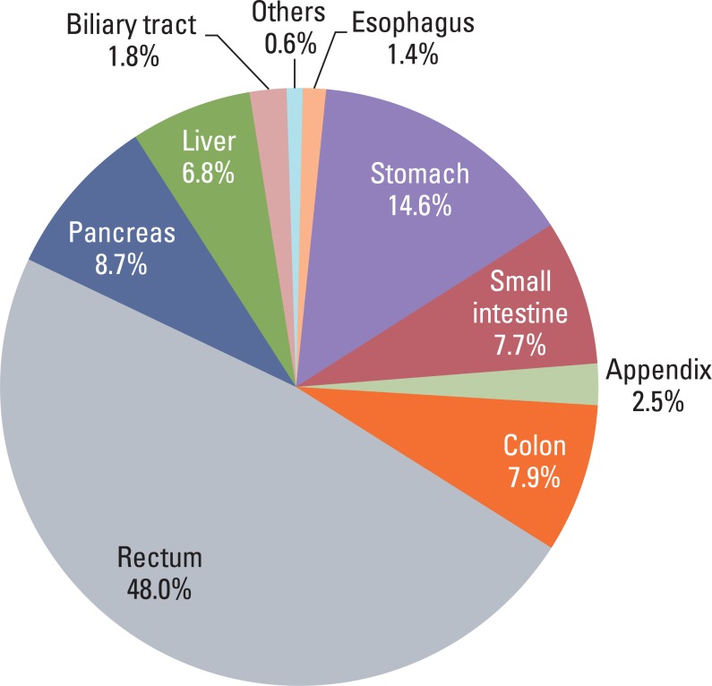
We collected 4,951 pathology reports of patients with GEP-NET. The mean age of patients was 54.66 years old and the sex ratio (M : F) was 1.5 : 1. The highest incidence of GEP-NET was observed in the rectum (48%), while the lowest incidence, 1.4%, was observed in the esophagus. A summary of data identified in the collected pathology reports is shown in Table 1. There was no description of the extension or size if the tumor was obtained by biopsy or partial excision. The rate of lymph node evaluation was low because most tumors were well-differentiated endocrine tumor (WDET) and were treated by simple tumor resection without lymph node dissection. Because some tumors were reported as carcinoid according to the 1980 WHO classification, we reclassified tumors according to the criteria of 2000 WHO classification if data of tumor size and extension were available; WDET, well-differentiated endocrine carcinoma (WDEC), poorly differentiated endocrine carcinoma/small cell carcinoma (PDEC), mixed exocrine-endocrine carcinoma (MEEC), and metastatic endocrine carcinoma (MEC). We also classified tumors according to the 2010 WHO classification if information on mitosis or Ki-67 labeling index were available.
2. Distribution of 4951 GEP-NET according to the organ system
Most GEP-NET were found in the rectum or the stomach (Fig. 1) and the most common site was the rectum. Multiplicity was found in 55 cases (1.3%) and was common in the rectum and stomach. Seventy three cases (1.5%) showed other concurrent pathology in either the same organ or another site: 56 adenocarcinomas in the same organ (stomach [n=34], rectum [n=7], colon [n=5], pancreas [n=4], appendix [n=2], small intestine [n=2], and biliary tree [n=2]), 7 adenomas in the same organ (colon [n=4], rectum [n=2], and stomach [n=1]), 4 lung cancer, 2 cervical cancer, 1 neurofibromatosis type I, 1 neurofibroma in the same organ, 1 retroperitoneal schwannoma, and 1 renal cell carcinoma.
3. Distribution of GEP-NET according to the 2000 and 2010 WHO classification
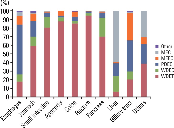
The distribution of tumors according to diagnosis is shown in Fig. 2. In the reclassification of the tumors according to the 2000 WHO classification, 601 WDET tumors were reclassified as WDEC. The proportion of GEP-NET according to the 2000 WHO classification in each site is shown in Fig. 3. Most rectal tumors were WDET, and most esophageal and biliary tumors were carcinoma.
4. Changes in the incidence of GEP-NET in Korea during the last decade
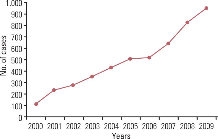
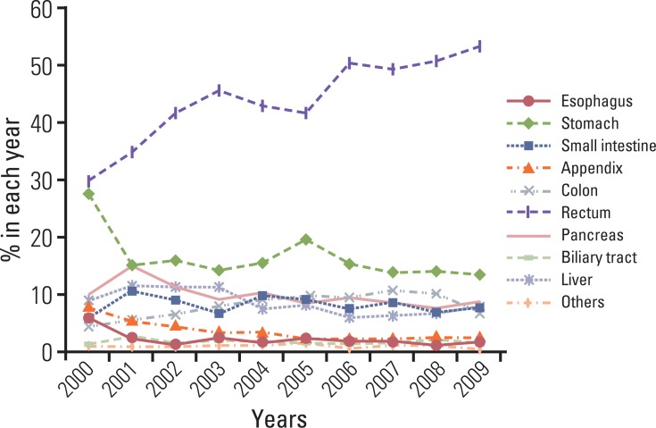
A continuous increase has been observed in the incidence of GEP-NET, with the incidence in 2009 being nine times that reported in 2000 (Fig. 4). During the last decade, the incidence of GEP-NET has been on the rise, particularly in the rectum (Fig. 5). Although changes in the incidence of GEP-NET according to the 2000 WHO classification was most significant in WDET, the incidence of WDEC and PDEC has also increased since 2003, though the incidence is still low in comparison with other malignancies of the digestive tract (Fig. 6).
5. Proportion of disease related death (DRD) in GEP-NETs according to the tumor site
Survival rate according to site is shown in Fig. 7. DRD was most common in the hepatobiliary system (62.2%), and least common in the rectum (5.1%). Most patients with rectal NET survived.
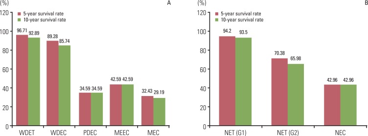
6. Five- and 10-year survival rate according to the 2000 and 2010 WHO classification of NET
The five- and 10-year survival rates of well differentiated tumors were distinctly different from those of poorly differentiated and metastatic tumors (Fig. 8). Five-year survival rates were similar to 10-year survival rates in each diagnosis group.
7. Prognostic significance of pathological parameters
For all cases examined, the site (p=0.000), diagnosis by 2000 WHO classification (p=0.000), extent (p=0.000), size (p=0.000), mitosis (p=0.000), Ki-67 labeling index (p=0.000), synaptophysin expression (p=0.003), lymphovascular invasion (p=0.000), perineural invasion (p=0.000), and lymph node metastasis (p=0.000) showed prognostic significance, however, chromogranin expression did not (p=0.148).
In the gastrointestinal tract, except for the appendix, all parameters, except synaptophysin (p=0.476) and chromogranin (p=0.089), showed prognostic significance. The lack of chromogranin expression appeared to be related to better survival, but it was not statistically significant. However, in the appendix, synaptophysin expression was associated with better prognosis, compared to synaptophysin negative tumors.
In analysis of WDET in all sites, chromogranin expression (p=0.018), lymphovascular invasion (p=0.024), lymph node metastasis (p=0.040) and size (p=0.024) showed prognostic significance, but mitosis (p=0.395), Ki-67 (p=0.817), synaptophysin (p=0.083), and perineural invasion (p=0.083) did not. However, in rectal WDET, synaptophysin expression and perineural invasion were significant prognostic factors. WDET in colon and rectal tumors showed the best prognosis, in contrast to the worst prognosis in biliary tract tumors.
In Kaplan-Meier survival analysis with Log-Rank test in WDEC, mitosis (p=0.002), Ki-67 (p=0.041), lymphovascular invasion (p=0.010), lymph node metastasis (p=0.033) & size (p=0.002) showed prognostic significance. The small intestinal tumor showed the best prognosis, in contrast to the worst prognosis in biliary tract tumors.
However, in the poorly differentiated neuroendocrine carcinoma, perineural invasion (p=0.011), tumor extension (p=0.000), and sites (p=0.007) showed prognostic significance. Gastric tumors showed the best prognosis, in contrast to the worst in biliary tract tumors. In the mixed exo- and endocrine tumor, lymph node metastasis (p=0.013), tumor extension (p=0.024), and sites (p=0.015) showed prognostic significance. Small intestinal and pancreatic tumors showed the best prognosis, incontrast to the worst prognosis in esophagus.
In this study, we described a significant increase in incidence of GEP-NET in Korea during the last decade. During the last decade, the incidence of GEP-NET in Korea has shown a significant increase. The rectal well differentiated NET was the most common and showed the most significant increase during the last decade, compared to the western report, which described the small intestinal NET as the most common. This change could be attributed to improved diagnostic tools, such as endoscopy. The incidence of small intestinal NET was 7.7%, which is similar to that reported in the Japanese study (9.6%) [16]. Incidence of WDEC, PDEC, MEEC, and MEC has shown a 5- to 9-fold increase in the last five years, although they are still relatively rare, compared to other malignant tumors of the digestive system. These changes are in concordance with the western report; however, the reasons are not yet clear.
Gosset and Masson first described carcinoids as endocrine-related tumors in 1914 [20]. Later, in 1963, Williams and Sandler classified them according to their embryologic site of origin as foregut, mid gut, and hind gut tumor [20]. Although it was the first description of morphological and biological differences between tumors, this classification did not reflect the diverse spectrum of GEP-NET. In the first WHO classification of GEP-NETS in 1980, the term carcinoid was used to describe all gastrointestinal neuroendocrine tumor regardless of biologic behavior and pathological characteristics [21]. In 2000 and 2004, the WHO reclassified NET into well-differentiated and poorly differentiated tumors [1]. Well-differentiated tumors were subdivided into benign, uncertain malignancy, or carcinoma. They recommended that the term carcinoid be used only for benign well-differentiated tumors, however, some pathologists are still using "carcinoid tumor" for all well-differentiated NET. The WHO recently updated the classification and proposed the term neuroendocrine neoplasms as the appropriate term for describing tumors originating from the diffuse endocrine system in the human body. According to the 2010 WHO classification, neuroendocrine neoplasms were devided into NET G1, NET G2, and neuroendocrine carcinoma NEC based on cell proliferation. In this retrospective study, the 2000 WHO classification system was used for diagnosis of GEP-NET. This is because all of the pathology reports we used were made before the 2010 WHO classification and mitotic counts were recorded in only 32.7% of collected pathology reports. Some tumors were diagnosed as carcinoid regardless of the size and extent according to the 1980 WHO classification. Therefore, all carcinoids were reclassified according to the 2000 WHO classification based on tumor size and extent, if the information was available, and 17.68% of carcinoids were then reclassified as well differentiated neuroendocrine carcinoma. This means that the classification system used for diagnosis should be mentioned in the pathology report.
In regard to the biologic behavior of GEP-NET, ICD-O3 classified the L-cell type carcinoid in rectum or appendix as uncertain malignancy coded as /1. This has recently caused some confusion in the category of malignancy of GEP-NET, particularly the small rectal WDET. According to the 2000 WHO classification, the rectal WDET, the most common tumor in Korea, belongs to the benign or uncertain malignancy. However, the WHO classification updated in 2010 recommended classification of all GEP-NET as malignancy, although the L-cell type rectal NET remains as uncertain malignancy. However, there is no definitive description of diagnostic criteria of L-cell type tumor. In 2011, a Colonoscopy Study Group of the Korean Society of Coloproctology described the metastatic potential of colorectal carcinoid [22]. In colorectal NET, lymph node metastasis was found in 5.1% (21 out of 414 cases) and was related to tumor size. The rectal NET measuring less than 10 mm had a 1.99% lymph node metastatic rate (7 out of 359 cases). However, they did not describe the association with patient's survival. Jernman et al. [23] found that none of the NET G1 had metastatic disease. In addition, tumor recurrence was found only in incomplete resection of NET G1 tumors. In the Japanese report, metastasis showed significant correlation with tumor size. Tumors less than 10 mm and confined to the submucosa without vessel invasion did not show any metastasis [24]. However, few cases of small rectal carcinoid (<10 mm) accompanied by multiple liver metastasis have been reported in the literature [25]. All reported cases showed poor prognosis if the patient had liver metastasis, even though the tumor size was very small. However, the pathological factors that can predict the metastasis in small sized NET remains to be clarified. In our data, the 10-year survival rate of WDET was 92.89%, in contrast to 85.74% in WDEC, and 34.59% in PDEC. Rectal WDET showed a 10-year survival rate of 98% and DRD was found in nine cases among 1,045 WDET with available follow-up data. Only four of these patients were treated by excision or surgical resection with a clear margin, therefore, DRD was actually 0.38%. Tumor size was less than 10 mm (range, 2.5 to 9 mm) for all four cases, and survival duration ranged from 3-23 months. From these results, the rectal WDET should not be classified as benign even if the size is less than 10 mm. On the other hand, because most tumors showed a benign clinical course, it is also difficult to classify as malignancy. Further study of pathological parameters associated with patient survival, especially in the small rectal WDET is required.
To predict the biological behavior of GEP-NET, several pathological features have been proposed by many investigators. For proper evaluation, the minimum data set for the pathology report has recently been proposed [3,9,10]. Tumor extension, histological differentiation, lymph node metastasis, lymphatic invasion, and perineural invasion of the tumor are well known as the default features for pathological evaluation of GEP-NET as well as other malignant tumors of the gastrointestinal tract. Unlike other gastrointestinal tract epithelial malignancy, site and size are known to be very important as prognostic markers in NET. As neuroendocrine markers, synaptophysin (a small vesicle associated marker) and chromogranin (a large secretory granule associated marker) were the mainstay. In addition, proliferation activity, assessed by mitotic count or Ki-67 immunostain, was described as a significant prognostic factor by a European group. In this study, we performed survival analysis according to the pathological parameters described above, however, multivariate analysis (e.g., Cox Proportional Hazard Regression) was not available due to missing values for the eligible variables in the pathology reports. All pathological parameters examined in this study, except for chromogranin expression, showed prognostic significance in all cases. However, the lack of chromogranin expression showed a significant association with better prognosis in WDET. In WDET, mitosis (p=0.395) and Ki-67 labeling index (p=0.817) were not prognostic factors. This was attributed to the lack of variation between cases in each group. The size of the tumor, particularly those greater than 1.5 cm, and proliferative activity showed an association with poor survival in WDEC. Therefore, for proper evaluation of GEP-NET, all pathological parameters examined in this study, including the immunostain for neuroendocrine markers as well as Ki-67 labeling index, should be mentioned in the pathology report. Further study of the correlation between pathological parameters and multivariate analysis is needed.
The incidence of GEP-NET in Korea has shown a remarkable increase during the last decade, however, the distribution of tumors in the digestive system differs from that of western reports. Although tumor size and extent are well known prognostic factors, DRD was rarely found in the very small rectal WDET. Further study and meticulous assessment of pathological parameters, including immunostain, are critical to understanding the biological behavior of tumors as well as predicting prognosis of patients with GEP-NET.
Acknowledgements
The research was supported in part by a Korean Cancer Association Novartis Research Grant.

Conflict of interest relevant to this article was not reported.

  • 1. Kloppel G, Perren A, Heitz PU. The gastroenteropancreatic neuroendocrine cell system and its tumors: the WHO classification. Ann N Y Acad Sci. 2004;1014:13–27. PMID: 15153416ArticlePubMed
  • 2. Kloppel G, Couvelard A, Perren A, Komminoth P, McNicol AM, Nilsson O, et al. ENETS Consensus Guidelines for the Standards of Care in Neuroendocrine Tumors: towards a standardized approach to the diagnosis of gastroenteropancreatic neuroendocrine tumors and their prognostic stratification. Neuroendocrinology. 2009;90:162–166. PMID: 19060454ArticlePubMed
  • 3. Klimstra DS, Modlin IR, Coppola D, Lloyd RV, Suster S. The pathologic classification of neuroendocrine tumors: a review of nomenclature, grading, and staging systems. Pancreas. 2010;39:707–712. PMID: 20664470ArticlePubMed
  • 4. Bosman FT, Carneiro F, Hruban RH, Theise ND. WHO classification of tumours of the digestive system. 2010. 4th ed. Lyon: International Agency for Research on Cancer.
  • 5. Kloppel G, Rindi G, Anlauf M, Perren A, Komminoth P. Site-specific biology and pathology of gastroenteropancreatic neuroendocrine tumors. Virchows Arch. 2007;451(Suppl 1):S9–S27. PMID: 17684761ArticlePubMed
  • 6. Boudreaux JP, Klimstra DS, Hassan MM, Woltering EA, Jensen RT, Goldsmith SJ, et al. The NANETS consensus guideline for the diagnosis and management of neuroendocrine tumors: well-differentiated neuroendocrine tumors of the jejunum, ileum, appendix, and cecum. Pancreas. 2010;39:753–766. PMID: 20664473ArticlePubMed
  • 7. Washington MK, Tang LH, Berlin J, Branton PA, Burgart LJ, Carter DK, et al. Protocol for the examination of specimens from patients with neuroendocrine tumors (carcinoid tumors) of the colon and rectum. Arch Pathol Lab Med. 2010;134:176–180. PMID: 20121603ArticlePubMed
  • 8. Edge SB, Compton CC. The American Joint Committee on Cancer: the 7th edition of the AJCC cancer staging manual and the future of TNM. Ann Surg Oncol. 2010;17:1471–1474. PMID: 20180029ArticlePubMed
  • 9. Klimstra DS, Modlin IR, Adsay NV, Chetty R, Deshpande V, Gonen M, et al. Pathology reporting of neuroendocrine tumors: application of the Delphic consensus process to the development of a minimum pathology data set. Am J Surg Pathol. 2010;34:300–313. PMID: 20118772ArticlePubMed
  • 10. Vinik AI, Woltering EA, Warner RR, Caplin M, O'Dorisio TM, Wiseman GA, et al. NANETS consensus guidelines for the diagnosis of neuroendocrine tumor. Pancreas. 2010;39:713–734. PMID: 20664471ArticlePubMed
  • 11. Rindi G, Kloppel G, Alhman H, Caplin M, Couvelard A, de Herder WW, et al. TNM staging of foregut (neuro)endocrine tumors: a consensus proposal including a grading system. Virchows Arch. 2006;449:395–401. PMID: 16967267ArticlePubMedPMC
  • 12. Fritz A, Percy C, Jack A, Shanmugaratnam S, Sobin L, Parkin DM, et al. International classification of diseases for oncology (ICD-O). 2000. 3rd ed. Geneva: World Health Organization.
  • 13. Maggard MA, O'Connell JB, Ko CY. Updated population-based review of carcinoid tumors. Ann Surg. 2004;240:117–122. PMID: 15213627ArticlePubMedPMC
  • 14. Garcia-Carbonero R, Capdevila J, Crespo-Herrero G, Diaz-Perez JA, Martinez Del Prado MP, Alonso Orduna V, et al. Incidence, patterns of care and prognostic factors for outcome of gastroenteropancreatic neuroendocrine tumors (GEP-NETs): results from the National Cancer Registry of Spain (RGETNE). Ann Oncol. 2010;21:1794–1803. PMID: 20139156ArticlePubMed
  • 15. Rothenstein J, Cleary SP, Pond GR, Dale D, Gallinger S, Moore MJ, et al. Neuroendocrine tumors of the gastrointestinal tract: a decade of experience at the Princess Margaret Hospital. Am J Clin Oncol. 2008;31:64–70. PMID: 18376230ArticlePubMed
  • 16. Ito T, Sasano H, Tanaka M, Osamura RY, Sasaki I, Kimura W, et al. Epidemiological study of gastroenteropancreatic neuroendocrine tumors in Japan. J Gastroenterol. 2010;45:234–243. PMID: 20058030ArticlePubMed
  • 17. Lee H, Choi J, An JS, Kim H, Shin BK, Kim A, et al. The clinicopathological characteristics of gastrointestinal neuroendocrine tumors: an analysis of 65 cases. Korean J Pathol. 2007;41:149–157.
  • 18. Chetty R. An overview of practical issues in the diagnosis of gastroenteropancreatic neuroendocrine pathology. Arch Pathol Lab Med. 2008;132:1285–1289. PMID: 18684027ArticlePubMed
  • 19. Edge SB, Byrd DR, Compton CC, Fritz AG, Greene FL, Trotti A. AJCC cancer staging manual. 2010. 7th ed. New York: Springer.
  • 20. Arnold R. Endocrine tumours of the gastrointestinal tract. Introduction: definition, historical aspects, classification, staging, prognosis and therapeutic options. Best Pract Res Clin Gastroenterol. 2005;19:491–505. PMID: 16183523ArticlePubMed
  • 21. Williams ED, Siebenmann RE, Sobin LH. Histological typing of endocrine tumours. 1980. Geneva: World Health Organization.
  • 22. Colonoscopy Study Group of Korean Society of ColoproctologyClinical characteristics of colorectal carcinoid tumors. J Korean Soc Coloproctol. 2011;27:17–20. PMID: 21431092ArticlePubMedPMC
  • 23. Jernman J, Valimaki MJ, Louhimo J, Haglund C, Arola J. The novel WHO 2010 classification for gastrointestinal neuroendocrine tumours correlates well with the metastatic potential of rectal neuroendocrine tumours. Neuroendocrinology. 2012;95:317–324. PMID: 22327359ArticlePubMed
  • 24. Hirata I, Akutagawa H, Nishikawa T, Yasumoto S, Sanomura M, Egashira Y, et al. Clinicopathological study of colorectal carcinoid tumor, focusing on the indication for endoscopic mucosal resection (EMR). Stormach Intest. 2005;40:182–193.
  • 25. Tabata H, Matsumoto T, Kuwano Y, Yao T, Iida M. Rectal carcinoid tumor 10 mm in diameter accompanied by multiple liver metastases, report of a case. Stormach Intest. 2005;40:215–220.
Fig. 1
Distribution of 4,951 gastroenteropancreatic neuroendocrine tumors according to the organ system.
crt-44-157-g001.jpg
Fig. 2
Distribution of gastroenteropancreatic neuroendocrine tumors according to diagnosis by 2000 and 2010 classification.WDET, well-differntiated endocrine tumor; WDEC, well-differentiated endocrine carcinoma; PDEC, poorly differentiated endocrine carcinoma/small cell carcinoma; MEEC, mixed exocrine-endocrine carcinoma; MEC, metastatic endocrine carcinoma; NET, neuroendocrine tumor; NEC, neuroendocrine carcinoma.
crt-44-157-g002.jpg
Fig. 3
Site distribution of gastroenteropancreatic neuroendocrine tumors in Korea according to the 2000 WHO classification. The most common tumor is WDET in rectum. WHO, World Health Organization; WDET, well-differntiated endocrine tumor; MEC, metastatic endocrine carcinoma, MEEC, mixed exocrine-endocrine carcinoma; PDEC, poorly differentiated endocrine carcinoma/small cell carcinoma; WDEC, well-differentiated endocrine carcinoma.
crt-44-157-g003.jpg
Fig. 4
Changes in incidence of gastroenteropancreatic neuroendocrine tumors in Korea during the last decade according to the number of annual diagnosis.
crt-44-157-g004.jpg
Fig. 5
Changes in annual incidence of gastroenteropancreatic neuroendocrine tumors in each site during the last decade.
crt-44-157-g005.jpg
Fig. 6
Changes in incidence of gastroenteropancreatic neuroendocrine tumors in Korea during the last decade according to the 2000 WHO classification of tumors. WHO, World Health Organization; WDET, well-differntiated endocrine tumor;WDEC, well-differentiated endocrine carcinoma; PDEC, poorly differentiated endocrine carcinoma/small cell carcinoma; MEEC, mixed exocrine-endocrine carcinoma; MEC, metastatic endocrine carcinoma.
crt-44-157-g006.jpg
Fig. 7
Site distribution of gastroenteropancreatic neuroendocrine tumors according to the survival. Mean survival time is the longest in stomach neuroendocrine tumor (154.42±7.74 months) and the shortest in biliary tract neuroendocrine tumors (44.32±6.02 months). Disease related death is most common in hepatobiliary tract.
crt-44-157-g007.jpg
Fig. 8
Five- and 10-year survival rate according to the 2000 (A) and 2010 WHO classification of NET (B). According to the 2000 WHO classification, the difference of 10 year survival rate between NET G1 and NET G2 is more distinct than the difference between WDET and WDEC. WHO, World Health Organization; NET, neuroendocrine tumor; WDET, well-differntiated endocrine tumor; WDEC, well-differentiated endocrine carcinoma; PDEC, poorly differentiated endocrine carcinoma/small cell carcinoma; MEEC, mixed exocrine-endocrine carcinoma; MEC, metastatic endocrine carcinoma; NEC, neuroendocrine carcinoma.
crt-44-157-g008.jpg
Table 1
Summary of information identified in the pathology reports
Cases

Recorded Result

No. % No. %
Tumor size (mm) 3,045 61.5 100
 0-5 844 27.7
 6-10 1,077 35.4
 11-15 459 15.1
 >15 665 21.8
Tumor extension 3,440 69.5
Distant metastasis 2,094 42.3 84 4.0
Lymph node metastasis 791 16.0 392 49.6
Mitosis 1,598 28.4 100.0
 <2/10 HPF 1,405 87.9
 2-20/10 HPF 126 7.9
 >20/10 HPF 67 4.2
Ki-67 IHC 637 12.9 100.0
 ≤2/10 HPF 457 71.7
 3-20/10 HPF 108 17.0
 >20/10 HPF 72 11.3
Synatophysin IHC 2,410 48.7 2,265a) 94.0
Chromogranin IHC 2,455 49.6 1,677a) 68.3
Lymphovascular invasion 2,312 46.7 505a) 21.8
Perineural invasion 1,128 22.8 178a) 15.8
Survival 3,312 66.9 568b) 17.1

HPF, high power field; IHC, immunohistochemistry. a)No. of positive cases, b)Disease related death.

Figure & Data

REFERENCES

    Citations

    Citations to this article as recorded by  
    • Risk Factor Analysis of Lymph Node Metastasis for Rectal Neuroendocrine Tumors: Who Needs a Radical Resection in Rectal Neuroendocrine Tumors Sized 1–2 cm?
      Jin Sun Choi, Min Jung Kim, Rumi Shin, Ji Won Park, Seung Chul Heo, Seung-Yong Jeong, Kyu Joo Park, Seung-Bum Ryoo
      Annals of Surgical Oncology.2024; 31(4): 2414.     CrossRef
    • Somatostatin receptor 2 (SSTR2) expression is associated with better clinical outcome and prognosis in rectal neuroendocrine tumors
      Joo Young Kim, Jisup Kim, Yong-il Kim, Dong-Hoon Yang, Changhoon Yoo, In Ja Park, Baek-Yeol Ryoo, Jin-Sook Ryu, Seung-Mo Hong
      Scientific Reports.2024;[Epub]     CrossRef
    • Endoscopic mucosal resection using anchored snare Tip-in versus precut technique for small rectal neuroendocrine tumors
      Seung Wook Hong, Dong-Hoon Yang, Yoo Jin Lee, Dong Hoon Baek, Jaeyoung Chun, Hyun Gun Kim, Sung Joo Kim, Seung-Mo Hong, Dae-Seong Myung
      The Korean Journal of Internal Medicine.2024; 39(2): 238.     CrossRef
    • Assessing risk stratification in long-term outcomes of rectal neuroendocrine tumors following endoscopic resection: a multicenter retrospective study
      Hyun Jin Lee, Yun Seo, Chang Kyo Oh, Ji Min Lee, Hyun Ho Choi, Tae-Geun Gweon, Sung-Hak Lee, Dae Young Cheung, Jin Il Kim, Soo-Heon Park, Han Hee Lee
      Scandinavian Journal of Gastroenterology.2024; 59(7): 868.     CrossRef
    • Clinicopathological Characteristics, Survival and Prognostic Factors in Gastrointestinal Large Cell Neuroendocrine Carcinoma
      Lele Chang, Xuemei Zhang, Jiaxin Li, Qingwei Li
      American Journal of Clinical Oncology.2024; 47(8): 363.     CrossRef
    • Comparison of endoscopic resection therapies for rectal neuroendocrine tumors
      Meijiao Lu, Hongxia Cui, Mingjie Qian, Yating Shen, Jianhong Zhu
      Minimally Invasive Therapy & Allied Technologies.2024; 33(4): 207.     CrossRef
    • Klinische Bedeutung von neuroendokrinen Tumoren
      Philipp E. Hartrampf, Sebastian E. Serfling, Takahiro Higuchi, Jörg Bojunga, Alexander Weich, Rudolf A. Werner
      Die Radiologie.2024; 64(7): 536.     CrossRef
    • Mixed neuroendocrine–non-neuroendocrine neoplasm of the bile duct with long-term prognosis after neoadjuvant chemotherapy
      Shinya Nakamura, Masahiro Serikawa, Yasutaka Ishii, Yumiko Tatsukawa, Juri Ikemoto, Sayaka Miyamoto, Kenichiro Uemura, Shinya Takahashi, Koji Arihiro, Shiro Oka
      Clinical Journal of Gastroenterology.2024; 17(4): 717.     CrossRef
    • Advancements in Neuroendocrine Neoplasms: Imaging and Future Frontiers
      Luigi Asmundo, Francesco Rizzetto, Michael Blake, Mark Anderson, Amirkasra Mojtahed, William Bradley, Anuradha Shenoy-Bhangle, Carlos Fernandez-del Castillo, Motaz Qadan, Cristina Ferrone, Jeffrey Clark, Valentina Ambrosini, Maria Picchio, Paola Mapelli,
      Journal of Clinical Medicine.2024; 13(11): 3281.     CrossRef
    • Primary coexisting adenocarcinoma of the colon and neuroendocrine tumor of the duodenum: A case report and review of the literature
      Song Fei, Wei-Dong Wu, Han-Shuo Zhang, Shao-Jie Liu, Dan Li, Bo Jin
      World Journal of Gastrointestinal Surgery.2024; 16(8): 2724.     CrossRef
    • Pancreatic neuroendocrine tumors: tailoring imaging to specific clinical scenarios
      Katerina S. Konstantinoff, Ajaykumar C. Morani, Thomas A. Hope, Priya R. Bhosale, Isaac R. Francis, Motoyo Yano, Amir Iravani, Nikolaos A. Trikalinos, Malak Itani
      Abdominal Radiology.2023; 48(5): 1843.     CrossRef
    • Is lymphatic invasion of microrectal neuroendocrine tumors an incidental event?: A case report
      Jing-Xue Ran, Liang-Bi Xu, Wan-Wei Chen, Hao-Yi Yang, Yan Weng, Yong-Mei Peng
      World Journal of Clinical Cases.2023; 11(4): 859.     CrossRef
    • Comparison of Metastatic Patterns Among Neuroendocrine Tumors, Neuroendocrine Carcinomas, and Nonneuroendocrine Carcinomas of Various Primary Organs
      Hyung Kyu Park, Ghee Young Kwon
      Journal of Korean Medical Science.2023;[Epub]     CrossRef
    • Somatostatin receptor 2 in 10 different types of human non-neoplastic gastrointestinal neuroendocrine cells
      Hirofumi Watanabe, Fumiyoshi Fujishima, Michiaki Unno, Hironobu Sasano, Takashi Suzuki
      Pathology - Research and Practice.2023; 244: 154418.     CrossRef
    • Different miRNAs Related to FBXW7 Mutations or High Mitotic Indices Contribute to Rectal Neuroendocrine Tumors: A Pilot Study
      Ho Suk Kang, Ha Young Park, Hyun Lim, Il Tae Son, Min-Jeong Kim, Nan Young Kim, Min Jeong Kim, Eun Sook Nam, Seong Jin Cho, Mi Jung Kwon
      International Journal of Molecular Sciences.2023; 24(7): 6329.     CrossRef
    • High-Grade Pure Esophageal Neuroendocrine Carcinoma Arising From Barrett’s Mucosa: A Rare Phenomenon
      Parth Patel, Eli A Zaher, Hasan Sqour
      Cureus.2023;[Epub]     CrossRef
    • Clinicopathologic Impact of Peptide Hormonal Expression in Rectal Neuroendocrine Tumors
      Jisup Kim, Dong-Hoon Yang, HaeSung Jung, HyungJun Cho, Hyeung-Jin Jang, Changhoon Yoo, In Ja Park, Baek-Yeol Ryoo, Jin-Sook Ryu, Seung-Mo Hong
      Archives of Pathology & Laboratory Medicine.2023; 147(7): 797.     CrossRef
    • Understanding the increasing incidence of neuroendocrine tumors
      Surabhi Pathak, Jason S. Starr, Thorvardur Halfdanarson, Mohamad Bassam Sonbol
      Expert Review of Endocrinology & Metabolism.2023; 18(5): 377.     CrossRef
    • Pancreatic Neuroendocrine Tumor (Pan-NET) Presented by Abdominal Pain: A Case Report and Literature Review
      Matteo Regolo, Nicolas Cardaci, Clara Salmeri, Alfredo Laudani, Michele Colaci, Massimo Ippolito, Fabio Motta, Salvatore Magrì, Stefanie Parisi, Alfredo Gaetano Torcitto, Lorenzo Malatino
      Journal of Clinical Medicine.2023; 12(20): 6617.     CrossRef
    • Clinical Value of Clip-and-Snare Assisted Endoscopic Submucosal Resection in Treatment of Rectal Neuroendocrine Tumors
      Xin-Tong Jiang, Yang Hu, Jian Gong, Shi-Bin Guo
      Visceral Medicine.2023; 39(5): 140.     CrossRef
    • Clinicopathological characteristics of extrahepatic biliary neuroendocrine neoplasms in the gallbladder, extrahepatic biliary tract, and ampulla of Vater: A single-center cross-sectional study
      Young Mok Park, Hyung Il Seo, Byeong Gwan Noh, Suk Kim, Seung Baek Hong, Nam Kyung Lee, Dong Uk Kim, Sung Yong Han
      Annals of Hepato-Biliary-Pancreatic Surgery.2023; 27(4): 380.     CrossRef
    • Rectal Neuroendocrine Neoplasms: Why Is There a Global Variation?
      Jack Cope, Raj Srirajaskanthan
      Current Oncology Reports.2022; 24(3): 257.     CrossRef
    • Clinicopathologic Characteristics of Gastroenteropancreatic Neuroendocrine Tumors: Experience of a National Referral Hospital in Indonesia
      Marini Stephanie, Nur Rahadiani, Hasan Maulahela, Ridho Ardhi Syaiful, Diah Rini Handjari, Ening Krisnuhoni
      Open Access Macedonian Journal of Medical Sciences.2022; 10(A): 101.     CrossRef
    • Update on Epidemiology, Diagnosis, and Biomarkers in Gastroenteropancreatic Neuroendocrine Neoplasms
      Daisuke Takayanagi, Hourin Cho, Erika Machida, Atsushi Kawamura, Atsuo Takashima, Satoshi Wada, Takuya Tsunoda, Takashi Kohno, Kouya Shiraishi
      Cancers.2022; 14(5): 1119.     CrossRef
    • Neuroendocrine Tumors: a Relevant Clinical Update
      Emma N. Rizen, Alexandria T. Phan
      Current Oncology Reports.2022; 24(6): 703.     CrossRef
    • A small, low-grade rectal neuroendocrine tumor with lateral pelvic lymph node metastasis: a case report
      Seonhui Shin, Young-In Maeng, Seyun Jung, Chun-Seok Yang
      Annals of Coloproctology.2022; 38(4): 327.     CrossRef
    • Rectal neuroendocrine neoplasms: what the radiologists should know
      Mayur Virarkar, Dheeraj R. Gopireddy, Ajaykumar C. Morani, Ahmad Alkhasawneh, Sergio Piotr Klimkowski, Sindhu Kumar, Chandana Lall, Priya Bhosale
      Abdominal Radiology.2022; 47(12): 4016.     CrossRef
    • Risk Factors for Lymph Node Metastasis and Oncologic Outcomes in Small Rectal Neuroendocrine Tumors with Lymphovascular Invasion
      Seung-Joo Nam, Byung Chang Kim, Hee Jin Chang, Han Ho Jeon, Junho Kim, Su Young Kim
      Gut and Liver.2022; 16(2): 228.     CrossRef
    • Clinical related factors to neuroendocrine tumors in Ecuadorian patients: a logistic biplot approach
      Karime Montes Escobar, José Luis Vicente Villardón, Daniel Fabricio Alarcón Cano, Aline Siteneski
      Investigación Clínica.2022; 63(1): 19.     CrossRef
    • Clinicopathological Characteristics of Gastroenteropancreatic Neuroendocrine Tumors
      Mustafa Şentürk, Berkan Acar, Mehmet Aykut Yildirim, Murat Çakir, Tevfik Küçükkartallar, Celalettin Vatansev
      Pancreas.2022; 51(2): 159.     CrossRef
    • Epidemiology of neuroendocrine neoplasmas in Japan: based on analysis of hospital-based cancer registry data, 2009 – 2015
      Tomonobu Koizumi, Kengo Otsuki, Yuriko Tanaka, Shintaro Kanda
      BMC Endocrine Disorders.2022;[Epub]     CrossRef
    • State of the Art in Endoscopic Therapy for the Management of Gastroenteropancreatic Neuroendocrine Tumors
      Apostolis Papaefthymiou, Faidon-Marios Laskaratos, Apostolos Koffas, Anastasios Manolakis, Paraskevas Gkolfakis, Sergio Coda, Mikael Sodergren, Noriko Suzuki, Christos Toumpanakis
      Current Treatment Options in Oncology.2022; 23(7): 1014.     CrossRef
    • Clinical characteristics, prognostic factors, and survival trends in esophageal neuroendocrine carcinomas: A population‐based study
      Chuyan Chen, Haiyi Hu, Zhibo Zheng, Yi Yang, Wei Chen, Xinwei Qiao, Peng Li, Shutian Zhang
      Cancer Medicine.2022; 11(24): 4935.     CrossRef
    • Underwater endoscopic mucosal resection for rectal neuroendocrine tumors (with videos): a single center retrospective study
      Haitao Shi, Chuying Wang, Jie Wu, Bin Qin, Jiong Jiang, Na Liu, Yahua Song, Yun Qin, Shiyang Ma
      BMC Gastroenterology.2022;[Epub]     CrossRef
    • Endoscopic treatment for rectal neuroendocrine tumor: which method is better?
      Seung Min Hong, Dong Hoon Baek
      Clinical Endoscopy.2022; 55(4): 496.     CrossRef
    • Clinical outcomes of gastroduodenal neuroendocrine tumors according to their WHO grade: A single-institutional retrospective analysis
      Dae Gon Ryu, Su Jin Kim, Cheol Woong Choi, Dae Hwan Kang, Hyung Wook Kim, Su Bum Park, Hyeong Seok Nam, Si Hak Lee, Sun Hwi Hwang
      Medicine.2022; 101(37): e30397.     CrossRef
    • Establishment and validation of a clinicopathological prognosis model of gastroenteropancreatic neuroendocrine carcinomas
      Jing Chen, Yibing Liu, Ke Xu, Fei Ren, Bowen Li, Hong Sun
      Frontiers in Oncology.2022;[Epub]     CrossRef
    • Incidence and survival of neuroendocrine neoplasia in England 1995–2018: A retrospective, population-based study
      Benjamin E. White, Brian Rous, Kandiah Chandrakumaran, Kwok Wong, Catherine Bouvier, Mieke Van Hemelrijck, Gincy George, Beth Russell, Rajaventhan Srirajaskanthan, John K. Ramage
      The Lancet Regional Health - Europe.2022; 23: 100510.     CrossRef
    • The risk and prognostic factors for G1 pancreatic neuroendocrine tumors: A retrospective analysis of the SEER database
      Zhengqi Wu, Xiaotong Qiu, Yao Zhi, Xiaoju Shi, Guoyue Lv
      Frontiers in Oncology.2022;[Epub]     CrossRef
    • MODERN PATHOLOGICAL CONSTELLATION FOR GASTROENTEROPANCREATIC NEUROENDOCRINE TUMORS AND IMMUNOHISTOCHEMICAL PANEL
      P. Vladova
      Trakia Journal of Sciences.2022; 20(4): 346.     CrossRef
    • An esophageal submucosal tumor treated with submucosal tunneling endoscopic resection: an unexpected result
      Ming-Yan Cai, Mariana Ferreira Cardoso, Yan Zhu, Yun-Shi Zhong, Ping-Hong Zhou
      Gastroenterology Report.2021; 9(3): 269.     CrossRef
    • A systematic review of chromogranin A (CgA) and its biomedical applications, unveiling its structure-related functions
      Kyuhyung Choi, Manhyuk Han, Seung Joong Kim
      Journal of the Korean Physical Society.2021; 78(5): 427.     CrossRef
    • Current Paradigm of Treatment for Pancreatic Neuroendocrine Tumor
      Min Je Sung, Moon Jae Chung
      The Korean Journal of Pancreas and Biliary Tract.2021; 26(1): 24.     CrossRef
    • Epidemiology, Incidence, and Prevalence of Neuroendocrine Neoplasms: Are There Global Differences?
      Satya Das, Arvind Dasari
      Current Oncology Reports.2021;[Epub]     CrossRef
    • Targeted Cancer Therapy: What’s New in the Field of Neuroendocrine Neoplasms?
      Anna La Salvia, Paula Espinosa-Olarte, Maria Del Carmen Riesco-Martinez, Beatriz Anton-Pascual, Rocío Garcia-Carbonero
      Cancers.2021; 13(7): 1701.     CrossRef
    • Systemic Treatment of Advanced Gastroenteropancreatic Neuroendocrine Tumors in Korea: Literature Review and Expert Opinion
      Changhoon Yoo, Chung Ryul Oh, Seung-Tae Kim, Woo Kyun Bae, Hye-Jin Choi, Do-Youn Oh, Myung-Ah Lee, Baek-Yeol Ryoo
      Cancer Research and Treatment.2021; 53(2): 291.     CrossRef
    • Clinicopathological characteristics and prognosis of 232 patients with poorly differentiated gastric neuroendocrine neoplasms
      Deng Han, Yuan-Liang Li, Zhi-Wei Zhou, Fei Yin, Jie Chen, Fang Liu, Yan-Fen Shi, Wei Wang, Yu Zhang, Xian-Jun Yu, Jian-Ming Xu, Run-Xiang Yang, Chao Tian, Jie Luo, Huang-Ying Tan
      World Journal of Gastroenterology.2021; 27(21): 2895.     CrossRef
    • Standardization of the pathologic diagnosis of appendiceal mucinous neoplasms
      Dong-Wook Kang, Baek-hui Kim, Joon Mee Kim, Jihun Kim, Hee Jin Chang, Mee Soo Chang, Jin-Hee Sohn, Mee-Yon Cho, So-Young Jin, Hee Kyung Chang, Hye Seung Han, Jung Yeon Kim, Hee Sung Kim, Do Youn Park, Ha Young Park, So Jeong Lee, Wonae Lee, Hye Seung Lee,
      Journal of Pathology and Translational Medicine.2021; 55(4): 247.     CrossRef
    • Multi-institutional development and validation of a nomogram to predict recurrence after curative resection of gastric neuroendocrine/mixed adenoneuroendocrine carcinoma
      Hualong Zheng, YaJun Zhao, Qingliang He, Hankun Hao, Yantao Tian, Bingbing Zou, Lixin Jiang, Xiantu Qiu, Yanbing Zhou, Zhi Li, Yanchang Xu, Gang Zhao, Fangqin Xue, Shuliang Li, Weihua Fu, Yongxiang Li, Xiaojun Zhou, Yong Li, Zhenggang Zhu, Jinping Chen, Z
      Gastric Cancer.2021; 24(2): 503.     CrossRef
    • Comparative study of laparoscopic versus open radical gastrectomy in advanced gastric neuroendocrine carcinoma: Analysis from a high-volume institution
      Ya-Jun Zhao, Lv-Ping Zhuang, Yi-Yang Liu, Ren-Cheng Chen, Jun Zhang, Hai-Xing Zhu, Yong-Cang Wang, Hao You, Ming-Wei Zhang, Xin-Yang He
      Asian Journal of Surgery.2020; 43(3): 488.     CrossRef
    • Potential Pitfalls in Diagnostic Digital Image Analysis: Experience with Ki-67 and PHH3 in Gastrointestinal Neuroendocrine Tumors
      Sean M Hacking, Sujata Sajjan, Lili Lee, Yonah Ziemba, Mallorie Angert, Yihe Yang, Cao Jin, Hector Chavarria, Nidhi Kataria, Swachi Jain, Mansoor Nasim
      Pathology - Research and Practice.2020; 216(3): 152753.     CrossRef
    • Imaging and Contrast-enhanced Ultrasound features of duodenal neuroendocrine tumor: A case report
      Yan-Qun Zhang, Jie-Xian Wen, Rong-Kui Luo, Hai-Xia Yuan, Wen-Ping Wang
      Clinical Hemorheology and Microcirculation.2020; 76(1): 27.     CrossRef
    • Rare cancers are not rare in Asia as well: The rare cancer burden in East Asia
      Tomohiro Matsuda, Young-Joo Won, RuRu Chun-ju Chiang, Jiwon Lim, Kumiko Saika, Keisuke Fukui, Wen-Chung Lee, Laura Botta, Alice Bernasconi, Annalisa Trama
      Cancer Epidemiology.2020; 67: 101702.     CrossRef
    • Islet Tumors
      Tiffany J. Sinclair, George A. Poultsides
      Advances in Surgery.2020; 54: 69.     CrossRef
    • Primary Large Cell Neuroendocrine Carcinoma of the Esophagus Disguised as a Food Impaction
      Harsh D. Patel, Jared Beck, Archish Kataria, Glenn W. Gross, Juan Echavarria
      ACG Case Reports Journal.2020; 7(6): e00384.     CrossRef
    • Pancreatic nodule positive for 68-Ga-DOTAPEPTIDE-PET: NET or ectopic spleen? The importance of a good differential diagnosis
      Francesco Serra, Gianluca De Matteis, Sara Delfanti, Monica Feltri, Alessandro Vanoli, Giorgio Cavenaghi, Anna Gallotti, Marcello Maestri, Silvia Brugnatelli
      Clinical and Translational Imaging.2020; 8(5): 307.     CrossRef
    • Gastric neuroendocrine tumor with Helicobacter pylori-associated chronic gastritis
      Giovanni Serio, Armando Dell’Anna, Anna Debenedittis, Elisa Stasi, Maria Arcangela Cascarano
      International Journal of Surgery Case Reports.2020; 75: 361.     CrossRef
    • Chromogranin A Expression in Rectal Neuroendocrine Tumors Is Associated With More Aggressive Clinical Behavior and a Poorer Prognosis
      Jisup Kim, Joo Young Kim, Eun Hye Oh, Changhoon Yoo, In Ja Park, Dong-Hoon Yang, Baek-Yeol Ryoo, Jin-Sook Ryu, Seung-Mo Hong
      American Journal of Surgical Pathology.2020; 44(11): 1496.     CrossRef
    • Clinicopathological characteristics and prognosis of 77 cases with type 3 gastric neuroendocrine tumours
      Yuan-Liang Li, Xu-Dong Qiu, Jie Chen, Yu Zhang, Jie Li, Jian-Ming Xu, Chao Wang, Zhi-Rong Qi, Jie Luo, Huang-Ying Tan
      World Journal of Gastrointestinal Oncology.2020; 12(12): 1416.     CrossRef
    • Clinicopathological Study of 100 Cases of Neuroendocrine Neoplasms of the Gastroenteropancreatic System: A Tertiary Cancer Center Experience
      Satarupa Tarak Nath Samanta, Shailee Paresh Bhai Mehta, Trupti S Patel, Dhaval H Jetly
      Indian Journal of Medical and Paediatric Oncology.2020; 41(03): 340.     CrossRef
    • Validation and proposed modification of the 8th edition American Joint Committee on Cancer staging system for patients with esophageal neuroendocrine neoplasms: Evaluation of a revised lymph node classification
      Haihong Wang, Yaobing Chen, Guoliang Pi, Ying Zhu, Shengli Yang, Hong Mei, Zhenyu Lin, Tao Zhang
      Oncology Letters.2020;[Epub]     CrossRef
    • Efficacy and safety of lanreotide in Korean patients with metastatic, well-differentiated gastroenteropancreatic-neuroendocrine tumors: a retrospective analysis
      Junho Kang, Changhoon Yoo, Hee-Sang Hwang, Seung-Mo Hong, Kyu-pyo Kim, Sun Young Kim, Yong-Sang Hong, Tae Won Kim, Baek-Yeol Ryoo
      Investigational New Drugs.2019; 37(4): 763.     CrossRef
    • A rare rarity: Neuroendocrine tumor of the esophagus
      Elisa Giannetta, Valentina Guarnotta, Francesca Rota, Federica de Cicco, Federica Grillo, Annamaria Colao, Antongiulio Faggiano
      Critical Reviews in Oncology/Hematology.2019; 137: 92.     CrossRef
    • Preoperative diagnosis of well‐differentiated neuroendocrine tumor in common hepatic duct by brush cytology: A case report
      Jiwoon Choi, Kyong Joo Lee, Sung Hoon Kim, Mee‐Yon Cho
      Diagnostic Cytopathology.2019; 47(7): 720.     CrossRef
    • Recent Updates in the Management of Advanced Pancreatic Neuroendocrine Tumors
      Chang Min Cho
      The Korean Journal of Gastroenterology.2019; 73(3): 124.     CrossRef
    • Clinical significance of programmed death 1/programmed death ligand 1 pathway in gastric neuroendocrine carcinomas
      Min-Wei Yang, Xue-Liang Fu, Yong-Sheng Jiang, Xiao-Jing Chen, Ling-Ye Tao, Jian-Yu Yang, Yan-Miao Huo, Wei Liu, Jun-Feng Zhang, Pei-Feng Liu, Qiang Liu, Rong Hua, Zhi-Gang Zhang, Yong-Wei Sun, De-Jun Liu
      World Journal of Gastroenterology.2019; 25(14): 1684.     CrossRef
    • Endoscopic Ultrasound-Guided Radiofrequency Ablation: A New Therapeutic Approach for Pancreatic Neuroendocrine Tumors
      Kira Oleinikov, Alain Dancour, Julia Epshtein, Ariel Benson, Haggi Mazeh, Ilanit Tal, Shay Matalon, Carlos A Benbassat, Dan M Livovsky, Eran Goldin, David J Gross, Harold Jacob, Simona Grozinsky-Glasberg
      The Journal of Clinical Endocrinology & Metabolism.2019; 104(7): 2637.     CrossRef
    • Clinical analysis of 547 patients with neuroendocrine tumors in a Chinese population: A single‐center study
      Lijie Song, Xuejia Zhai, Shunli Yu, Yihui Ma, Feng Wang, Xuxu Yu, Shuang Tao, Yujin Lian, Minjie Yang, Weili Tao, Qingxia Fan
      Cancer Medicine.2019; 8(8): 3729.     CrossRef
    • Endoscopic, transanal, laparoscopic, and transabdominal management of rectal neuroendocrine tumors
      Louis de Mestier, Diane Lorenzo, Caroline Fine, Jérôme Cros, Olivia Hentic, Thomas Walter, Yves Panis, Anne Couvelard, Guillaume Cadiot, Philippe Ruszniewski
      Best Practice & Research Clinical Endocrinology & Metabolism.2019; 33(5): 101293.     CrossRef
    • Intrapancreatic accessory spleen false positive to 68Ga-Dotatoc: case report and literature review
      Francesco Lancellotti, Luca Sacco, Saverio Cerasari, Vittoria Bellato, Simone Cicconi, Antonio Ciardi, Edoardo Maria Muttillo, Tiziana Feola, Roberto Caronna, Piero Chirletti
      World Journal of Surgical Oncology.2019;[Epub]     CrossRef
    • Novel Finding of Paired Box 5 (PAX5) Cytoplasmic Staining in Well-differentiated Rectal Neuroendocrine Tumors (Carcinoids) and Its Diagnostic and Potentially Prognostic Utility
      Zhiyan Fu, Chunlai Zuo, Christine E. Sheehan, Deepa T. Patil, Jingmei Lin, Zhaohai Yang, Hwajeong Lee
      Applied Immunohistochemistry & Molecular Morphology.2019; 27(6): 454.     CrossRef
    • Duodenal neuroendocrine tumors
      A. V. Alekberzade, N. N. Krylov, T. N. Garmanova, R. Shahbazov, F. Azari, K. S. Zuykova, E. D. Litovchenko
      Khirurgiya. Zhurnal im. N.I. Pirogova.2019; (7): 87.     CrossRef
    • [68Ga]-Dota Peptide PET/CT in Neuroendocrine Tumors: Main Clinical Applications
      Elisabetta Giovannini, Giampiero Giovacchini, Elisa Borsò, Patrizia Lazzeri, Mattia Riondato, Rossella Leoncini, Valerio Duce, Andrea Ciarmiello
      Current Radiopharmaceuticals.2019; 12(1): 11.     CrossRef
    • Multifocal G1-G2 gastric neuroendocrine tumors: Differentiating between Type I, II and III, a clinicopathologic review
      Khaled Algashaamy, Monica Garcia-Buitrago
      World Journal of Clinical Cases.2019; 7(17): 2413.     CrossRef
    • Establishment and characterization of a novel neuroendocrine carcinoma cell line derived from a human ascending colon tumor
      Seiichi Shinji, Norihiko Sasaki, Takeshi Yamada, Michihiro Koizumi, Ryo Ohta, Akihisa Matsuda, Yasuyuki Yokoyama, Goro Takahashi, Masahiro Hotta, Keisuke Hara, Kohki Takeda, Koji Ueda, Sho Kuriyama, Toshiyuki Ishiwata, Yoshibumi Ueda, Takashi Murakami, Yo
      Cancer Science.2019; 110(12): 3708.     CrossRef
    • Gastric neuroendocrine tumors
      A. V. Alekberzade, N. N. Krylov, E. M. Lipnitskiy, R. O. Shakhbazov, F. Azari
      Khirurgiya. Zhurnal im. N.I. Pirogova.2019; (12): 111.     CrossRef
    • Resectability of Rectal Neuroendocrine Tumors Using Endoscopic Mucosal Resection with a Ligation Band Device and Endoscopic Submucosal Dissection
      Hong Kyu Lim, Seong Jun Lee, Dong Hoon Baek, Do Youn Park, Bong Eun Lee, Eun Young Park, Joon Woo Park, Gwang Ha Kim, Geun Am Song
      Gastroenterology Research and Practice.2019; 2019: 1.     CrossRef
    • Epidemiological trends of neuroendocrine tumours over three decades in Queensland, Australia
      David Wyld, Mark H. Wan, Julie Moore, Nathan Dunn, Philippa Youl
      Cancer Epidemiology.2019; 63: 101598.     CrossRef
    • Lymphovascular invasion as a prognostic value in small rectal neuroendocrine tumor treated by local excision: A systematic review and meta-analysis
      Ho Suk Kang, Mi Jung Kwon, Tae-Hwan Kim, Junhee Han, Young-Su Ju
      Pathology - Research and Practice.2019; 215(11): 152642.     CrossRef
    • Gastroenteropancreatic Neuroendocrine Tumor with Hepatic Metastasis Misdiagnosed as Hepatocellular Carcinoma
      Seul Ki Kim, Sang Chul Kim, Seok Gyo Seo, Hong Joo Kim, Yong Kyun Cho, Woo Kyu Jeon, Byung Ik Kim
      The Korean Journal of Medicine.2019; 94(5): 449.     CrossRef
    • Clinicopathologic Characteristics and Survival of Patients with Gastroenteropancreatic Neuroendocrine Neoplasm in a Multi-Ethnic Asian Institution
      Wai Meng Tai, Sze Huey Tan, Damien Meng Yew Tan, Kelvin Siu Hoong Loke, David Chee Eng Ng, Sean Xuexian Yan, Jacqueline Siok Gek Hwang, Kiat Hon Lim, Lih Ming Loh, Peng Chin Kek, Brian Kim Poh Goh, Ser Yee Lee, Alexander Yaw Fui Chung, Simon Yew Kuang Ong
      Neuroendocrinology.2019; 108(4): 265.     CrossRef
    • Primary Esophagogastric Neuroendocrine Carcinoma: a Retrospective Study from the Nottingham Upper Gastrointestinal Cancer Center
      Constantinos Savva, Philip Kaye, Irshad Soomro, Simon L. Parsons, Eleanor James, Srinivasan Madhusudan
      Journal of Gastrointestinal Cancer.2018; 49(1): 85.     CrossRef
    • Transanal Endoscopic Microsurgery for Patients with Rare Rectal Tumors
      Xin Wu, Guole Lin, Huizhong Qiu, Jiaolin Zhou
      Journal of Laparoendoscopic & Advanced Surgical Techniques.2018; 28(5): 546.     CrossRef
    • A case of small intestinal neuroendocrine carcinoma diagnosed using double-balloon endoscopy with long-term survival
      Seiji Kawano, Yuichi Miyashima, Yoshio Miyabe, Yoshinari Kawai, Toshihiro Murata, Masashi Uda, Toshihiro Inokuchi, Hiroyuki Okada
      Clinical Journal of Gastroenterology.2018; 11(3): 240.     CrossRef
    • How should incidental NEN of the pancreas and gastrointestinal tract be followed?
      Riccardo Ariotti, Stefano Partelli, Francesca Muffatti, Valentina Andreasi, Francesca Della Sala, Massimo Falconi
      Reviews in Endocrine and Metabolic Disorders.2018; 19(2): 139.     CrossRef
    • Identification of Phosphohistone H3 Cutoff Values Corresponding to Original WHO Grades but Distinguishable in Well-Differentiated Gastrointestinal Neuroendocrine Tumors
      Min Jeong Kim, Mi Jung Kwon, Ho Suk Kang, Kyung Chan Choi, Eun Sook Nam, Seong Jin Cho, Hye-Rim Park, Soo Kee Min, Jinwon Seo, Ji-Young Choe, Hyoung-Chul Park
      BioMed Research International.2018; 2018: 1.     CrossRef
    • A meta-analysis of Prognostic factor of Pancreatic neuroendocrine neoplasms
      Yong Gao, Hao Gao, Guangfu Wang, Lingdi Yin, Wenbin Xu, Yunpeng Peng, Junli Wu, Kuirong Jiang, Yi Miao
      Scientific Reports.2018;[Epub]     CrossRef
    • Everolimus in the treatment of neuroendocrine tumors: efficacy, side-effects, resistance, and factors affecting its place in the treatment sequence
      Lingaku Lee, Tetsuhide Ito, Robert T. Jensen
      Expert Opinion on Pharmacotherapy.2018; 19(8): 909.     CrossRef
    • Pancreatic Neuroendocrine Neoplasms: A Role of Laparoscopy in Surgical Treatment: Review
      Jacek Szeliga, Marek Jackowski
      Surgical Laparoscopy, Endoscopy & Percutaneous Techniques.2018; 28(3): 147.     CrossRef
    • Clinicopathological features and outcome for neuroendocrine neoplasms of gastroesophageal junction: A population‐based study
      Panpan Zhang, Wei Wang, Ming Lu, Changqing Zeng, Jie Chen, Enxiao Li, Huangying Tan, Wei Wang, Xianjun Yu, Qiyun Tang, Jiemin Zhao, Lin Shen, Jie Li
      Cancer Medicine.2018; 7(9): 4361.     CrossRef
    • Neuroendocrine tumors with hepatic metastases: A review of evolving treatment options
      Steven D. Colquhoun
      Liver Research.2018; 2(2): 92.     CrossRef
    • Endoscopic Treatment Outcome of Rectal Neuroendocrine Tumors Removed by Ligation-assisted Endoscopic Submucosal Resection
      Min Geun Gu, Si Hyung Lee
      The Korean Journal of Gastroenterology.2018; 72(3): 128.     CrossRef
    • Clinicopathological features and prognosis of patients with gastric neuroendocrine tumors: A population‐based study
      Fang‐Xi Tian, Yu‐Qing Cai, Lv‐Ping Zhuang, Ming‐Fang Chen, Zhong‐Biao Xiu, Yi Zhang, Hong Liu, Zhi‐Hong Liu, Guo‐Ping Liu, Chen Zeng, Fei‐Ling Lin, Jing Liu, Si‐Ting Huang, Liang‐Zhi Zhang, Hua‐Yang Lin
      Cancer Medicine.2018; 7(11): 5359.     CrossRef
    • Global burden of neuroendocrine tumors and changing incidence in Kentucky
      Aman Chauhan, Qian Yu, Neha Ray, Zainab Farooqui, Bin Huang, Eric B. Durbin, Thomas Tucker, Mark Evers, Susanne Arnold, Lowell B. Anthony
      Oncotarget.2018; 9(27): 19245.     CrossRef
    • Gastroenteropancreatic neuroendocrine tumor registry study in China
      Lin Cong, Wenming Wu, Wenhui Lou, Jinwan Wang, Feng Gu, Jiaming Qian, Jianming Xu, Feng Bi, Sanjun Cai, Chengwei Tang, Yulong He, Jie Zhong, Yupei Zhao
      Journal of Pancreatology.2018; 1(1): 35.     CrossRef
    • Incidence and clinical characteristics of gastroenteropancreatic neuroendocrine tumor in Korea: a single-center experience
      Chul-Hyun Lim, In Seok Lee, Byoung Yeon Jun, Jin Su Kim, Yu Kyung Cho, Jae Myung Park, Sang Young Roh, Myung Ah Lee, Sang Woo Kim, Myung-Gyu Choi
      The Korean Journal of Internal Medicine.2017; 32(3): 452.     CrossRef
    • Uncommon Anal Neoplasms
      Amitesh C. Roy, David Wattchow, David Astill, Simron Singh, Susan Pendlebury, Kirsten Gormly, Eva Segelov
      Surgical Oncology Clinics of North America.2017; 26(1): 143.     CrossRef
    • Surveillance and comparison of surgical prognosis for asymptomatic and symptomatic non-functioning pancreatic neuroendocrine tumors
      Wenhao Ge, Dongkai Zhou, Shaoyan Xu, Weilin Wang, Shusen Zheng
      International Journal of Surgery.2017; 39: 127.     CrossRef
    • Recent updates on grading and classification of neuroendocrine tumors
      Joo Young Kim, Seung-Mo Hong, Jae Y. Ro
      Annals of Diagnostic Pathology.2017; 29: 11.     CrossRef
    • Mixed adenoneuroendocrine carcinoma derived from the cystic duct: A case report
      Yu-ki Takemoto, Tomoyuki Abe, Hironobu Amano, Keiji Hanada, Akihito Okazaki, Tomoyuki Minami, Tsuyoshi Kobayashi, Masahiro Nakahara, Shuji Yonehara, Hideki Ohdan, Toshio Noriyuki
      International Journal of Surgery Case Reports.2017; 39: 29.     CrossRef
    • Loss of Progesterone Receptor Expression Is an Early Tumorigenesis Event Associated with Tumor Progression and Shorter Survival in Pancreatic Neuroendocrine Tumor Patients
      Sung Joo Kim, Soyeon An, Jae Hoon Lee, Joo Young Kim, Ki-Byung Song, Dae Wook Hwang, Song Cheol Kim, Eunsil Yu, Seung-Mo Hong
      Journal of Pathology and Translational Medicine.2017; 51(4): 388.     CrossRef
    • Clinical outcomes of everolimus in patients with advanced, nonfunctioning pancreatic neuroendocrine tumors: a multicenter study in Korea
      Kyong Joo Lee, Jae Hee Cho, Sang Hyub Lee, Si Young Song, Kwang Hyuk Lee, Seok Jeong, Ji Kon Ryu, Sang Myung Woo, Seungmin Bang, Jong Kyun Lee, Tae Hoon Lee, Woo Hyun Paik, Yong Tae Kim, Woo Jin Lee
      Cancer Chemotherapy and Pharmacology.2017; 80(4): 799.     CrossRef
    • Clinicopathological, treatment, and prognosis study of 43 gastric neuroendocrine carcinomas
      De-Jun Liu, Xue-Liang Fu, Wei Liu, Lu-Ying Zheng, Jun-Feng Zhang, Yan-Miao Huo, Jiao Li, Rong Hua, Qiang Liu, Yong-Wei Sun
      World Journal of Gastroenterology.2017; 23(3): 516.     CrossRef
    • Is endoscopic ultrasonography essential for endoscopic resection of small rectal neuroendocrine tumors?
      Su Bum Park, Dong Jun Kim, Hyung Wook Kim, Cheol Woong Choi, Dae Hwan Kang, Su Jin Kim, Hyeong Seok Nam
      World Journal of Gastroenterology.2017; 23(11): 2037.     CrossRef
    • Predictive Factors for Lymph Node Metastasis and Prognostic Factors for Survival in Rectal Neuroendocrine Tumors
      Beonghoon Sohn, Yoomin Kwon, Seung-Bum Ryoo, Inho Song, Yoon-Hye Kwon, Dong Woon Lee, Sang Hui Moon, Ji Won Park, Seung-Yong Jeong, Kyu Joo Park
      Journal of Gastrointestinal Surgery.2017; 21(12): 2066.     CrossRef
    • Efficacy of Precut Endoscopic Mucosal Resection for Treatment of Rectal Neuroendocrine Tumors
      Hoonsub So, Su Hyun Yoo, Seungbong Han, Gwang-un Kim, Myeongsook Seo, Sung Wook Hwang, Dong-Hoon Yang, Jeong-Sik Byeon
      Clinical Endoscopy.2017; 50(6): 585.     CrossRef
    • Gastroenteropancreatic—origin neuroendocrine carcinomas
      Yong Gyun Won, Kyung-Jin Seo, Jiyeon Hyeon, Ok Ran Shin, Eundeok Chang, Der Sheng Sun, Hae Sung Won, Yoon Ho Ko, Sae Jung Na, Su Lim Lee, Young Mi Ku, Dong Soo Lee
      Medicine.2017; 96(49): e9009.     CrossRef
    • Sex comb on midleg like-2 is a novel specific marker for the diagnosis of gastroenteropancreatic neuroendocrine tumors
      Jiao-Jiao Yang, Hua Huang, Ming-Bing Xiao, Feng Jiang, Wen-Kai Ni, Yi-Fei Ji, Cui-Hua Lu, Run-Zhou Ni
      Experimental and Therapeutic Medicine.2017; 14(2): 1749.     CrossRef
    • A nation-wide retrospective epidemiological study of gastroenteropancreatic neuroendocrine neoplasms in china
      Jin-Hu Fan, Yu-Qing Zhang, Su-Sheng Shi, Yuan-Jia Chen, Xing-Hua Yuan, Li-Ming Jiang, Shao-Ming Wang, Li Ma, Yu-Tong He, Chang-Yan Feng, Xi-Bin Sun, Qing Liu, Katrina Deloso, Yihebali Chi, You-Lin Qiao
      Oncotarget.2017; 8(42): 71699.     CrossRef
    • Irinotecan plus cisplatin followed by octreotide long-acting release maintenance treatment in advanced gastroenteropancreatic neuroendocrine carcinoma: IPO-NEC study
      Jie Li, Ming Lu, Zhihao Lu, Zhongwu Li, Yiqiang Liu, Li Yang, Jian Li, Xiaotian Zhang, Jun Zhou, Xicheng Wang, Jifang Gong, Jing Gao, Yan Li, Lin Shen
      Oncotarget.2017; 8(15): 25669.     CrossRef
    • Cap-assisted EMR for rectal neuroendocrine tumors: comparisons with conventional EMR and endoscopic submucosal dissection (with videos)
      Dong-Hoon Yang, Yangsoon Park, Sang Hyoung Park, Kyung-Jo Kim, Byong Duk Ye, Jeong-Sik Byeon, Seung-Jae Myung, Suk-Kyun Yang
      Gastrointestinal Endoscopy.2016; 83(5): 1015.     CrossRef
    • Surgical Treatment as a Principle for Patients with High-Grade Pancreatic Neuroendocrine Carcinoma: A Nordic Multicenter Comparative Study
      Sven-Petter Haugvik, Eva Tiensuu Janson, Pia Österlund, Seppo W. Langer, Ragnhild Sørum Falk, Knut Jørgen Labori, Lene Weber Vestermark, Henning Grønbæk, Ivar Prydz Gladhaug, Halfdan Sorbye
      Annals of Surgical Oncology.2016; 23(5): 1721.     CrossRef
    • Evaluation of the risk factors associated with rectal neuroendocrine tumors: a big data analytic study from a health screening center
      Jeung Hui Pyo, Sung Noh Hong, Byung-Hoon Min, Jun Haeng Lee, Dong Kyung Chang, Poong-Lyul Rhee, Jae Jun Kim, Sun Kyu Choi, Sin-Ho Jung, Hee Jung Son, Young-Ho Kim
      Journal of Gastroenterology.2016; 51(12): 1112.     CrossRef
    • Recent Updates on Neuroendocrine Tumors From the Gastrointestinal and Pancreatobiliary Tracts
      Joo Young Kim, Seung-Mo Hong
      Archives of Pathology & Laboratory Medicine.2016; 140(5): 437.     CrossRef
    • Metronomic temozolomide as second line treatment for metastatic poorly differentiated pancreatic neuroendocrine carcinoma
      C. De Divitiis, C. von Arx, A. M. Grimaldi, D. Cicala, F. Tatangelo, A. Arcella, G. M. Romano, E. Simeone, R. V. Iaffaioli, P. A. Ascierto, S. Tafuto
      Journal of Translational Medicine.2016;[Epub]     CrossRef
    • Epidemiological features of gastroenteropancreatic neuroendocrine tumors in Chengdu city with a population of 14 million based on data from a single institution
      Lin-Jie Guo, Chun-Hui Wang, Cheng-Wei Tang
      Asia-Pacific Journal of Clinical Oncology.2016; 12(3): 284.     CrossRef
    • The Clinicopathologic Features and Treatment of 607 Hindgut Neuroendocrine Tumor (NET) Patients at a Single Institution
      Seung Tae Kim, Sang Yun Ha, Jeeyun Lee, Sung No Hong, Dong Kyung Chang, Young Ho Kim, Yoon Ah Park, Jung Wook Huh, Yong Beom Cho, Seong Hyeon Yun, Woo Yong Lee, Hee Cheol Kim, Young Suk Park
      Medicine.2016; 95(19): e3534.     CrossRef
    • Clinicopathological Profile of Pure Neuroendocrine Neoplasms of the Esophagus: A South Indian Center Experience
      Govind Babu Kanakasetty, Loknatha Dasappa, Kuntegowdanahalli Chinnagiriyappa Lakshmaiah, Mangesh Kamath, Linu Abraham Jacob, Suresh Babu Mallekavu, Lakkavalli Krishnappa Rajeev, Rudresha Antapura Haleshappa, Lokesh Kadabur Nagendrappa, Smitha Carol Saldan
      Journal of Oncology.2016; 2016: 1.     CrossRef
    • Endoscopic treatments for rectal neuroendocrine tumors smaller than 16 mm: a meta-analysis
      Hai-Ping Zhang, Wei Wu, Sheng Yang, Jun Lin
      Scandinavian Journal of Gastroenterology.2016; 51(11): 1345.     CrossRef
    • Lymph node size is not a reliable criterion for predicting nodal metastasis in rectal neuroendocrine tumours
      B. C. Kim, Y. E. Kim, H. J. Chang, S. H. Lee, E. G. Youk, D.‐S. Lee, J. B. Lee, E.‐J. Lee, M. J. Kim, D. K. Sohn, J. H. Oh
      Colorectal Disease.2016;[Epub]     CrossRef
    • Prognostic Validity of the American Joint Committee on Cancer and the European Neuroendocrine Tumors Staging Classifications for Pancreatic Neuroendocrine Tumors
      Jae Hee Cho, Ji Kon Ryu, Si Young Song, Jin-Hyeok Hwang, Dong Ki Lee, Sang Myung Woo, Young-Eun Joo, Seok Jeong, Seung-Ok Lee, Byung Kyu Park, Young Koog Cheon, Jimin Han, Tae Nyeun Kim, Jun Kyu Lee, Sung-Hoon Moon, Hyunjin Kim, Eun Taek Park, Jae Chul Hw
      Pancreas.2016; 45(7): 941.     CrossRef
    • Endoscopic Resection for Small Rectal Neuroendocrine Tumors: Comparison of Endoscopic Submucosal Resection with Band Ligation and Endoscopic Submucosal Dissection
      Byoung Wook Bang, Jin Seok Park, Hyung Kil Kim, Yong Woon Shin, Kye Sook Kwon, Joon Mee Kim
      Gastroenterology Research and Practice.2016; 2016: 1.     CrossRef
    • Surgical treatment and prognosis of gastric neuroendocrine neoplasms: a single-center experience
      Chaoyong Shen, Huijiao Chen, Haining Chen, Yuan Yin, Luyin Han, Jiaju Chen, Sumin Tang, Xiaonan Yin, Zongguang Zhou, Bo Zhang, Zhixin Chen
      BMC Gastroenterology.2016;[Epub]     CrossRef
    • The clinicopathological significance of angiogenesis in hindgut neuroendocrine tumors obtained via an endoscopic procedure
      Yoichiro Okubo, Osamu Motohashi, Norisuke Nakayama, Ken Nishimura, Rika Kasajima, Yohei Miyagi, Manabu Shiozawa, Emi Yoshioka, Masaki Suzuki, Kota Washimi, Kae Kawachi, Madoka Nito, Yoichi Kameda, Tomoyuki Yokose
      Diagnostic Pathology.2016;[Epub]     CrossRef
    • A Case of Endoscopically Complete Remission of Esophageal Neuroendocrine Tumors by Concurrent Chemoradiation Therapy
      Myung Hee Kim, Hyun Yong Jeong, Jae Kyu Seong, Hee Seok Moon, Sun Hyung Kang, Duk Ki Kim
      The Korean Journal of Gastroenterology.2016; 68(5): 265.     CrossRef
    • Outcomes after local excision for rectal neuroendocrine tumor
      Yoomin Kwon, Seung-Bum Ryoo, Inho Song, Yoon-Hye Kwon, Dong Woon Lee, Sang Hui Moon, Ji Won Park, Seung-Yong Jeong, Kyu Joo Park
      Korean Journal of Clinical Oncology.2016; 12(2): 104.     CrossRef
    • Multivariate analysis of metastasis-related risk factors for patients with gastroenteropancreatic neuroendocrine tumors based on clinicopathological and endoscopic features
      Caiyun Tang, Lingqi Gong, Wenli Zou, Jie Zhang, Yuqian Zhou, Xiaoping Wu, Fanggen Lu, Chunhui Ouyang, Xiaowei Liu
      Oncology Reports.2016; 36(6): 3343.     CrossRef
    • Management of gastric and duodenal neuroendocrine tumors
      Yuichi Sato, Satoru Hashimoto, Ken-ichi Mizuno, Manabu Takeuchi, Shuji Terai
      World Journal of Gastroenterology.2016; 22(30): 6817.     CrossRef
    • Surgical management for non-functional pancreatic neuroendocrine neoplasms with synchronous liver metastasis: A consensus from the Chinese Study Group for Neuroendocrine Tumors (CSNET)
      Kaizhou Jin, Jin Xu, Jie Chen, Minhu Chen, Rufu Chen, Ye Chen, Zhiyu Chen, Bin Cheng, Yihebali Chi, Shi-Ting Feng, Deliang Fu, Baohua Hou, Dan Huang, Heguang Huang, Qiang Huang, Jie Li, Ying Li, Houjie Liang, Rong Lin, An'an Liu, Jixi Liu, Xubao Liu, Ming
      International Journal of Oncology.2016; 49(5): 1991.     CrossRef
    • ENETS Consensus Guidelines Update for the Management of Patients with Functional Pancreatic Neuroendocrine Tumors and Non-Functional Pancreatic Neuroendocrine Tumors
      M. Falconi, B. Eriksson, G. Kaltsas, D.K. Bartsch, J. Capdevila, M. Caplin, B. Kos-Kudla, D. Kwekkeboom, G. Rindi, G. Klöppel, N. Reed, R. Kianmanesh, R.T. Jensen
      Neuroendocrinology.2016; 103(2): 153.     CrossRef
    • Epidemiological trends of pancreatic and gastrointestinal neuroendocrine tumors in Japan: a nationwide survey analysis
      Tetsuhide Ito, Hisato Igarashi, Kazuhiko Nakamura, Hironobu Sasano, Takuji Okusaka, Koji Takano, Izumi Komoto, Masao Tanaka, Masayuki Imamura, Robert T. Jensen, Ryoichi Takayanagi, Akira Shimatsu
      Journal of Gastroenterology.2015; 50(1): 58.     CrossRef
    • Neuroendocrine neoplasms of the extrahepatic bile duct: radiologic and clinical characteristics
      Nurhee Hong, Hyoung Jung Kim, Jae Ho Byun, So Yeon Kim, Kyoung Won Kim, Jin Hee Kim, Seung-Mo Hong
      Abdominal Imaging.2015; 40(1): 181.     CrossRef
    • Circumferential submucosal incision prior to endoscopic mucosal resection provides comparable clinical outcomes to submucosal dissection for well-differentiated neuroendocrine tumors of the rectum
      Dae Young Cheung, Soo Kyoung Choi, Hyung-Keun Kim, Sung Soo Kim, Hiun-Suk Chae, Kyung Jin Seo, Young-Seok Cho
      Surgical Endoscopy.2015; 29(6): 1500.     CrossRef
    • Non-L-cell Immunophenotype and Large Tumor Size in Rectal Neuroendocrine Tumors Are Associated With Aggressive Clinical Behavior and Worse Prognosis
      Joo Young Kim, Ki-Suk Kim, Kyung-Jo Kim, In Ja Park, Jong Lyul Lee, Seung-Jae Myung, Yangsoon Park, Young Soo Park, Chang Sik Yu, Jin Cheon Kim, Eunsil Yu, Hyeung-Jin Jang, Seung-Mo Hong
      American Journal of Surgical Pathology.2015; 39(5): 632.     CrossRef
    • Aggressive Locoregional Treatment Improves the Outcome of Liver Metastases from Grade 3 Gastroenteropancreatic Neuroendocrine Tumors
      Shunda Du, Jianjiao Ni, Linqian Weng, Fei Ma, Shaohua Li, Wenze Wang, Xinting Sang, Xin Lu, Shouxian Zhong, Yilei Mao
      Medicine.2015; 94(34): e1429.     CrossRef
    • Gastroenteropancreatic Neuroendocrine Tumors with Liver Metastases in Korea: A Clinicopathological Analysis of 72 Cases in a Single Institute
      Yooju Shin, Sang Yun Ha, Jiyeon Hyeon, Boram Lee, Jeeyun Lee, Kee-Taek Jang, Kyoung-Mee Kim, Young Suk Park, Cheol-Keun Park
      Cancer Research and Treatment.2015; 47(4): 738.     CrossRef
    • Prognostic Significance of Defining L-Cell Type on the Biologic Behavior of Rectal Neuroendocrine Tumors in Relation with Pathological Parameters
      Jin Hee Sohn, Mee-Yon Cho, Yangsoon Park, Hyunki Kim, Woo Ho Kim, Joon Mee Kim, Eun Sun Jung, Kyoung-Mee Kim, Jae Hyuk Lee, Hee Kyung Chan, Do Youn Park, Mee Joo, Sujin Kim, Woo Sung Moon, Mi Seon Kang, So-Young Jin, Yun Kyung Kang, Sun Och Yoon, HyeSeung
      Cancer Research and Treatment.2015; 47(4): 813.     CrossRef
    • Neuroendocrine Carcinomas of the Gastroenteropancreatic System: A Comprehensive Review
      Emma Ilett, Seppo Langer, Ingrid Olsen, Birgitte Federspiel, Andreas Kjær, Ulrich Knigge
      Diagnostics.2015; 5(2): 119.     CrossRef
    • A Re-evaluation of Colorectal Neuroendocrine Tumors Based on WHO 2010
      Akira Watanabe, Mitsuyoshi Ota, Yusuke Suwa, Shinsuke Suzuki, Atsushi Ishibe, Jun Watanabe, Kazuteru Watanabe, Yasushi Ichikawa, Kingo Hirasawa, Mikiko Tanabe, Itaru Endo
      Nippon Daicho Komonbyo Gakkai Zasshi.2015; 68(2): 61.     CrossRef
    • Gastroenteropancreatic high‐grade neuroendocrine carcinoma
      Halfdan Sorbye, Jonathan Strosberg, Eric Baudin, David S. Klimstra, James C. Yao
      Cancer.2014; 120(18): 2814.     CrossRef
    • Mixed Large Cell Neuroendocrine Carcinoma and Adenocarcinoma with Spindle Cell and Clear Cell Features in the Extrahepatic Bile Duct
      John Wysocki, Rishi Agarwal, Laura Bratton, Jeremy Nguyen, Mandy Crause Weidenhaft, Nathan Shores, Hillary Z. Kimbrell
      Case Reports in Pathology.2014; 2014: 1.     CrossRef
    • Spectrum of Gastroenteropancreatic NENs in Routine Histological Examinations of Bioptic and Surgical Specimen: A Study of 161 Cases Collected from 17 Departments of Pathology in the Czech Republic
      Václav Mandys, Tomáš Jirásek
      Gastroenterology Research and Practice.2014; 2014: 1.     CrossRef
    • Italian Association of Clinical Endocrinologists (AME) position statement: a stepwise clinical approach to the diagnosis of gastroenteropancreatic neuroendocrine neoplasms
      Franco Grimaldi, Nicola Fazio, Roberto Attanasio, Andrea Frasoldati, Enrico Papini, Francesco Angelini, Roberto Baldelli, Debora Berretti, Sara Bianchetti, Giancarlo Bizzarri, Marco Caputo, Roberto Castello, Nadia Cremonini, Anna Crescenzi, Maria Vittoria
      Journal of Endocrinological Investigation.2014; 37(9): 875.     CrossRef
    • Clinical features and management of type I gastric carcinoids
      Yuichi Sato
      Clinical Journal of Gastroenterology.2014; 7(5): 381.     CrossRef
    • Diagnosis and Treatment of Rectal Neuroendocrine Tumor
      Hyun Deok Shin
      Korean Journal of Medicine.2014; 87(4): 415.     CrossRef
    • Clinical, pathological and prognostic characteristics of gastroenteropancreatic neuroendocrine neoplasms in China: a retrospective study
      Xianbin Zhang, Li Ma, Haidong Bao, Jing Zhang, Zhongyu Wang, Peng Gong
      BMC Endocrine Disorders.2014;[Epub]     CrossRef
    • The clinical features and treatment modality of esophageal neuroendocrine tumors: a multicenter study in Korea
      Chang Geun Lee, Yun Jeong Lim, Seun Ja Park, Byung Ik Jang, Seok Reyol Choi, Jae Kwang Kim, Yong-Tae Kim, Joo Young Cho, Chang Hun Yang, Hoon Jai Chun, Si Young Song
      BMC Cancer.2014;[Epub]     CrossRef
    • A Modified Duodenal Neuroendocrine Tumor Staging Schema Better Defines the Risk of Lymph Node Metastasis and Disease-free Survival
      Swapnil D. Kachare, Kendall R. Liner, Nasreen A. Vohra, Emmanuel E. Zervos, Timothy L. Fitzgerald
      The American Surgeon™.2014; 80(8): 821.     CrossRef
    • A Case of Small Cell Neuroendocrine Tumor Occurring at Hilar Bile Duct
      Bum Chul Kim, Tae Jun Song, Hyuk Lee, Mee Joo, Won Ki Bae, Nam-Hoon Kim, Kyung-Ah Kim, June Sung Lee
      The Korean Journal of Gastroenterology.2013; 62(5): 301.     CrossRef
    • Neuroendocrine Tumors
      Young Suk Park
      Korean Journal of Medicine.2013; 85(4): 354.     CrossRef
    • Role of radiotherapy for pancreatobiliary neuroendocrine tumors
      Jeongshim Lee, Jinhyun Choi, Chihwan Choi, Jinsil Seong
      Radiation Oncology Journal.2013; 31(3): 125.     CrossRef
    • Proposal for a Standardized Pathology Report of Gastroenteropancreatic Neuroendocrine Tumors: Prognostic Significance of Pathological Parameters
      Mee-Yon Cho, Jin Hee Sohn, So Young Jin, Hyunki Kim, Eun Sun Jung, Mi-Jung Kim, Kyoung-Mee Kim, Woo Ho Kim, Joon Mee Kim, Yun Kyung Kang, Joon Hyuk Choi, Dae Young Kang, Youn Wha Kim, Eun Hee Choi
      Korean Journal of Pathology.2013; 47(3): 227.     CrossRef
    • Multiple Neuroendocrine Tumor of the Distal Ileum
      Seung Min Lee, Jong Hoon Lee
      The Korean Journal of Gastroenterology.2013; 61(2): 110.     CrossRef
    • The Epidemiology of Neuroendocrine Tumors in Taiwan: A Nation-Wide Cancer Registry-Based Study
      Hui-Jen Tsai, Chun-Chieh Wu, Chia-Rung Tsai, Sheng-Fung Lin, Li-Tzong Chen, Jeffrey S. Chang, Olga Y. Gorlova
      PLoS ONE.2013; 8(4): e62487.     CrossRef
    • Which endoscopic treatment is the best for small rectal carcinoid tumors?
      Hyun Ho Choi
      World Journal of Gastrointestinal Endoscopy.2013; 5(10): 487.     CrossRef
    • Prognostic impact of p16 and p21 on gastroenteropancreatic neuroendocrine tumors
      SHUZHENG LIU, YUXI CHANG, JIE MA, XU LI, XIAOHONG LI, JINHU FAN, RONG HUANG, GUANGCAI DUAN, XIBIN SUN
      Oncology Letters.2013; 6(6): 1641.     CrossRef
    • Epidemiology of gastroenteropancreatic neuroendocrine tumours
      M. Fraenkel, M.K. Kim, A. Faggiano, G.D. Valk
      Best Practice & Research Clinical Gastroenterology.2012; 26(6): 691.     CrossRef

    • PubReader PubReader
    • ePub LinkePub Link
    • Cite
      CITE
      export Copy Download
      Close
      Download Citation
      Download a citation file in RIS format that can be imported by all major citation management software, including EndNote, ProCite, RefWorks, and Reference Manager.

      Format:
      • RIS — For EndNote, ProCite, RefWorks, and most other reference management software
      • BibTeX — For JabRef, BibDesk, and other BibTeX-specific software
      Include:
      • Citation for the content below
      Current Trends of the Incidence and Pathological Diagnosis of Gastroenteropancreatic Neuroendocrine Tumors (GEP-NETs) in Korea 2000-2009: Multicenter Study
      Cancer Res Treat. 2012;44(3):157-165.   Published online September 30, 2012
      Close
    • XML DownloadXML Download
    Current Trends of the Incidence and Pathological Diagnosis of Gastroenteropancreatic Neuroendocrine Tumors (GEP-NETs) in Korea 2000-2009: Multicenter Study
    Image Image Image Image Image Image Image Image
    Fig. 1 Distribution of 4,951 gastroenteropancreatic neuroendocrine tumors according to the organ system.
    Fig. 2 Distribution of gastroenteropancreatic neuroendocrine tumors according to diagnosis by 2000 and 2010 classification.WDET, well-differntiated endocrine tumor; WDEC, well-differentiated endocrine carcinoma; PDEC, poorly differentiated endocrine carcinoma/small cell carcinoma; MEEC, mixed exocrine-endocrine carcinoma; MEC, metastatic endocrine carcinoma; NET, neuroendocrine tumor; NEC, neuroendocrine carcinoma.
    Fig. 3 Site distribution of gastroenteropancreatic neuroendocrine tumors in Korea according to the 2000 WHO classification. The most common tumor is WDET in rectum. WHO, World Health Organization; WDET, well-differntiated endocrine tumor; MEC, metastatic endocrine carcinoma, MEEC, mixed exocrine-endocrine carcinoma; PDEC, poorly differentiated endocrine carcinoma/small cell carcinoma; WDEC, well-differentiated endocrine carcinoma.
    Fig. 4 Changes in incidence of gastroenteropancreatic neuroendocrine tumors in Korea during the last decade according to the number of annual diagnosis.
    Fig. 5 Changes in annual incidence of gastroenteropancreatic neuroendocrine tumors in each site during the last decade.
    Fig. 6 Changes in incidence of gastroenteropancreatic neuroendocrine tumors in Korea during the last decade according to the 2000 WHO classification of tumors. WHO, World Health Organization; WDET, well-differntiated endocrine tumor;WDEC, well-differentiated endocrine carcinoma; PDEC, poorly differentiated endocrine carcinoma/small cell carcinoma; MEEC, mixed exocrine-endocrine carcinoma; MEC, metastatic endocrine carcinoma.
    Fig. 7 Site distribution of gastroenteropancreatic neuroendocrine tumors according to the survival. Mean survival time is the longest in stomach neuroendocrine tumor (154.42±7.74 months) and the shortest in biliary tract neuroendocrine tumors (44.32±6.02 months). Disease related death is most common in hepatobiliary tract.
    Fig. 8 Five- and 10-year survival rate according to the 2000 (A) and 2010 WHO classification of NET (B). According to the 2000 WHO classification, the difference of 10 year survival rate between NET G1 and NET G2 is more distinct than the difference between WDET and WDEC. WHO, World Health Organization; NET, neuroendocrine tumor; WDET, well-differntiated endocrine tumor; WDEC, well-differentiated endocrine carcinoma; PDEC, poorly differentiated endocrine carcinoma/small cell carcinoma; MEEC, mixed exocrine-endocrine carcinoma; MEC, metastatic endocrine carcinoma; NEC, neuroendocrine carcinoma.
    Current Trends of the Incidence and Pathological Diagnosis of Gastroenteropancreatic Neuroendocrine Tumors (GEP-NETs) in Korea 2000-2009: Multicenter Study
    Cases

    Recorded Result

    No. % No. %
    Tumor size (mm) 3,045 61.5 100
     0-5 844 27.7
     6-10 1,077 35.4
     11-15 459 15.1
     >15 665 21.8
    Tumor extension 3,440 69.5
    Distant metastasis 2,094 42.3 84 4.0
    Lymph node metastasis 791 16.0 392 49.6
    Mitosis 1,598 28.4 100.0
     <2/10 HPF 1,405 87.9
     2-20/10 HPF 126 7.9
     >20/10 HPF 67 4.2
    Ki-67 IHC 637 12.9 100.0
     ≤2/10 HPF 457 71.7
     3-20/10 HPF 108 17.0
     >20/10 HPF 72 11.3
    Synatophysin IHC 2,410 48.7 2,265a) 94.0
    Chromogranin IHC 2,455 49.6 1,677a) 68.3
    Lymphovascular invasion 2,312 46.7 505a) 21.8
    Perineural invasion 1,128 22.8 178a) 15.8
    Survival 3,312 66.9 568b) 17.1
    Table 1 Summary of information identified in the pathology reports

    HPF, high power field; IHC, immunohistochemistry. a)No. of positive cases, b)Disease related death.


    Cancer Res Treat : Cancer Research and Treatment
    Close layer
    TOP