Introduction
Childhood cancer survivors (CCSs) are at risk for impairment of gonadal function and infertility [1]. Pubertal development and progression should be monitored in adolescents of childhood cancer. Delayed puberty or stalled pubertal development could be signs of gonadal defect in adolescence [2]. Furthermore, gonadal and sexual function of young adult survivors of childhood cancer should be regularly evaluated because premature ovarian insufficiency (POI) can occur. POI is defined by persistent amenorrhea with elevated gonadotropin levels in duration occurring 5 years after diagnosis and before age of 40 [3]. Permanent loss of ovarian function within 5 years of cancer diagnosis is regarded as acute ovarian failure [4].
Children’s oncology group (COG) guideline recommends checking follicular stimulating hormone (FSH) and estradiol for patients suggestive of POI or desire information about potential for future fertility [5]. FSH is specific and sensitive to evaluate gonadal function as a screening tool of POI in female CCSs because defective ovaries can lead to increased serum gonadotropin levels [1]. However, the concentration of FSH varies depending on the period of menstruation. FSH might not be increased high enough in spite of gonadal dysfunction due to disruption of the hypothalamic-pituitary-ovarian axis after cranial irradiation.
Anti-müllerian hormone (AMH) is produced in granulosa cells of developing antral follicles. AMH regulates dominant follicle selection by decreasing the sensitivity of antral follicles to FSH [6]. Measuring AMH is of importance for evaluating ovarian reserve in adult women, especially when decreased future fertility is suspected. So far, age-specific reference values of AMH in adolescents and young adults (AYA) less than 25 years are not well established yet. Nevertheless, recent studies have reported that AMH levels have a peak after birth during mini-puberty followed by a decline [7,8]. A linear increase of AMH has been noted from 4 years to 8 years. Relatively steady concentrations of AMH have been observed from 8 years to 25 years, enabling us to expand the role of AMH in pediatric fields [7–9]. COG guideline also recommends consideration of AMH in patients treated with gonadotoxic treatments to assess for diminished ovarian reserve [5]. However, data of AMH concentrations in CCSs are limited [10,11]. Furthermore, POI is associated with increased cardiovascular disease risk and bone mineral density (BMD) is also liked with hypogonadism in general population [12,13]. So far, there has been no comprehensive study regarding gonadal function and associated health problems of CCSs in Korea. Thus, the aim of this study was to investigate gonadal function in CCSs and to evaluate health outcomes associated with gonadal function.
Materials and Methods
1. Subjects
Sixty-nine female CCSs who visited our out-patient clinic between August 2016 and July 2022 were enrolled. These subjects were diagnosed with cancer less than 20 years of age. Cancer therapy was completed at least 1 year prior to the last follow-up. They were treated with chemotherapeutic agent, surgery, and radiotherapy at the National Cancer Center. Subjects whose age was less than 15 years at the last follow-up were excluded from this study. Their baseline characteristics are shown in Table 1. Their mean age at diagnosis was 12.4±4.4 years (range, 0.4 to 19.8 years). Their median follow-up period was 10.8 years (range, 1 to 16.5 years). Their mean age at last follow-up was 24.0±4.7 years (range, 15.2 to 33.8 years). Diagnoses included leukemia, lymphoma, central nervous system (CNS) tumor, embryonal tumor, bone and soft tissue sarcoma, and ovarian tumor. Among survivors with CNS tumor, subjects with hypogonadotropic hypogonadism were ruled out from this study. Amenorrheic patients with normal FSH levels resulting from intracranial (midline) germ cell tumor were considered as having hypogonadotropic hypogonadism.
2. Methods
Medical records were retrospectively reviewed to obtain demographic and medical characteristics of CCSs. Endocrinologic evaluation such as thyroid function, sex hormones, and lipid profile were performed at the end of treatment, and usually, annually thereafter at the National Cancer Center. Gonadotropin releasing hormone agonist (GnRH agonist) was used during chemotherapy in female subjects who presented secondary sexual characteristics such as breast development, pubertal hair with sexual maturity rating (Tanner stages) 2 or more at diagnosis. The results of laboratory test (luteinizing hormone [LH], FSH) used in this study were conducted at least one year after completion of therapy and before sex hormone replacement therapy (HRT). Pubertal status, menstruation history, treatment history, and laboratory findings were obtained. Subjects were categorized into three groups with two serum FSH cutoff levels of 12 and 40 IU/L. FSH levels beyond 12 IU/L were regarded as abnormal [14]. Among subjects with FSH levels over 40 IU/L, absence of menstrual cycle at least 1 year after completion of cancer therapy was defined as POI in this study [15]. HRT was recommended and initiated in subjects with POI. HRT was transiently applied in some patients with secondary amenorrhea and levels of FSH between 12 and 40 IU/L according to the opinion of gynecologist.
Serum AMH level of less than 1 ng/mL was considered a low AMH level. Levels of LH and FSH were measured using a chemiluminescent immunoassay (ADVIA Centaur XP, Siemens, Erlangen, Germany). AMH levels were measured using an electrochemiluminescent immunoassay (Cobas 8000, Roche, Basel, Swiss). Total, high-density lipoprotein, and low-density lipoprotein (LDL) cholesterol levels were measured by enzymatic colorimetric methods using standard laboratory procedures (TBA-2000FR, Toshiba, Tokyo, Japan). Cumulative alkylating agents dose was calculated using the following equation [16]: cyclophosphamide equivalent dose (CED) (mg/m2)=1.0 (cumulative cyclophosphamide dose [mg/m2])+0.244 (cumulative ifosfamide dose [mg/m2])+0.857(cumulative procarbazine dose [mg/m2])+14.286 (cumulative chlorambucil dose [mg/m2])+15.0 (cumulative BCNU dose [mg/m2])+16.0 (cumulative CCNU dose [mg/m2])+40 (cumulative melphalan dose [mg/m2])+50 (cumulative Thio-TEPA dose [mg/m2])+100 (cumulative nitrogen mustard dose [mg/m2])+8.823 (cumulative busulfan dose [mg/m2]). BMD of the lumbar spine and femur was measured by dual energy X-ray absorptiometry (DXA; Horizon W, Hologic, Bedford, MA). BMD Z-score was calculated using Korean National Health and Nutrition Examination Survey (KNHANES) data [17].
3. Statistical analysis
All statistical analyses were performed using R Foundation for Statistical Computing ver. 4.1.2 (R Core Team (2021), R: A language and environment for statistical computing, Vienna, Austria). Kruskal Wallis test was performed to compare parameters including AMH levels among three groups according to FSH levels. Independent t test was used to compare data of age at diagnosis and body mass index (BMI) between two groups. Chi-squared test and Fisher’s exact test were used to analyze variables in groups according to FSH and AMH levels. Risk factors associated with low AMH levels were evaluated using logistic regression. Correlations between gonadotropin (FSH and LH), and AMH were analyzed using Pearson correlation. Paired t test was used to compare two AMH levels in the same patient. A p-value of less than 0.05 was considered statistically significant.
Results
1. Gonadal function of subjects
Median AMH level of subjects was 1.65 ng/mL (range, 0.02 to 12.5 ng/mL), which was measured at the age of 15–33 years (Table 1). Mean FSH concentrations of the three groups were 6.8±2.2, 20.7±7.9, and 88.7±26.3 IU/L, respectively. Of 69 subjects, 14 (20.3%) had POI, 14 (20.3%) had FSH levels between 12 and 40 IU/L. Forty-one of 69 subjects (59.4%) with normal FSH levels had less CED compared to the rest 28 subjects with abnormal FSH levels (p=0.008). Mean AMH levels in the three groups according to the FSH level were 4.1±3.0, 0.48±0.58, and 0.03±0.03 ng/mL, respectively (p < 0.001) (Fig. 1). All subjects with POI and 11 out of 14 patients (78.6%) with FSH levels between 12 and 40 IU/L had low AMH levels, whereas only three of 41 subjects (7.3%) with normal FSH levels had low AMH levels (p < 0.001) (Table 2). Six (42.9%) and 11 (78.6%) subjects of 14 with POI were treated with radiotherapy (pelvis or total body irradiation [TBI]) and stem cell transplantation (SCT), respectively (p=0.001 and p < 0.001, respectively) (Table 2). Three out of 14 with FSH levels between 12 and 40 IU/L had secondary amenorrhea. AMH levels were 0.4, 0.02, and 0.03 ng/mL in the three patients with secondary amenorrhea. Among 14 subjects with low AMH level and FSH below 40 IU/L, 13 subjects received more than 8,000 mg/m2 of CED of alkylating agents and three patients were treated with pelvic radiotherapy.
GnRH agonist was used in 31 of 69 patients. The mean age of patients with GnRH agonist was 15.2±2.4 years, while the mean age of patients without GnRH agonist was 10.1±4.4 (p < 0.001). The portion of patient with GnRH agonist was lower in subjects with POI than the other patients (p=0.037) (Table 2).
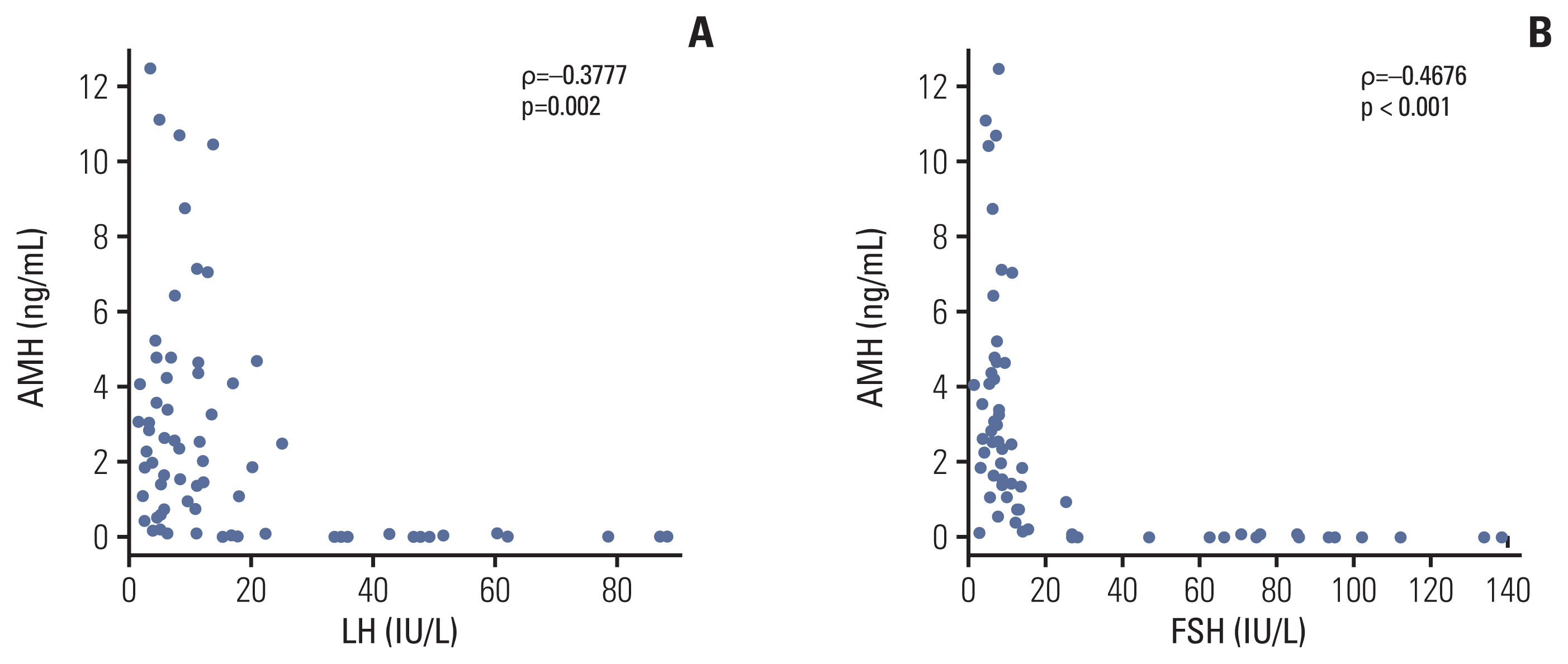
Of 21 CCSs with ovarian tumors, one patient had granulosa cell tumor and the rest of them had germ cell tumors. The mean AMH level was 3.53±3.11 ng/mL for those with ovarian tumors. Other patients had a mean AMH level of 2.10±2.91 ng/mL (p=0.083). Subjects with ovarian tumors were treated with less dose of CED than other patients (p < 0.001). No ovarian tumor patient underwent SCT or radiotherapy. Five of 21 ovarian tumors had FSH levels over 12 IU/L and 5 of 21 had low AMH levels. Only one patient received bilateral oophorectomy due to tumor recurrence, leading to POI and low AMH level (FSH, 75.2 IU/L; AMH, 0.08 ng/mL) and necessitating HRT. AMH levels were negatively correlated with LH (r=−0.3777, p=0.002) and FSH (r=−0.4679, p < 0.001) concentrations in 69 subjects (Fig. 2).
2. Risk factors for low AMH levels and follow-up AMH levels
Predictive factors for low AMH level were analyzed. Results are shown in Table 3. Age at diagnosis was not a significant risk factor for low AMH level in this study. CED, radiotherapy (pelvis or TBI), and SCT were significant predictive factors (odds ratios, 5.729, 16.0, and 19.5; p=0.002, p=0.011, and p < 0.001, respectively) in univariate analysis (Table 3). In multivariate analysis, CED and SCT were significant treatment factors for developing low AMH level (p=0.005 and p=0.002, respectively).
Follow-up AMH levels were measured for only 31 subjects. Mean AMH levels of 31 CCSs at age of 18.0 and 22.6 years were 2.16±3.44 and 2.80±3.61 ng/mL (p=0.076), respectively. Thirty-one CCSs were classified into two groups (low AMH vs. the others). AMH levels below 1 ng/mL at the last follow-up was defined as low AMH. Of 15 subjects with low AMH levels, the mean concentrations of AMH were 0.17±0.23 and 0.18±0.28 ng/mL at the age of 18.1±3.2 and 22.5±5.1 years, respectively (p=0.446) (Fig. 3). Of the rest 16 subjects, mean concentrations of AMH were 4.27±3.99 and 5.59±3.42 ng/mL at 17.9±2.4 and 22.8±3.6 years of age, respectively, indicating an increasing trend of AMH level over time (p < 0.001) (Fig. 3).
3. Lipid profile and BMD according to gonadal function
Lipid profile and BMD of the three groups according to FSH levels are shown in Table 4. The results of lipid profile were obtained in 68 of 69 subjects. Of three groups, BMI levels were not significantly different (p=0.576). Total and LDL cholesterols were significantly different in three groups according to FSH levels (p=0.047, p=0.030). Triglyceride were elevated in groups with FSH levels over 12 IU/L (p=0.045). DXA was performed in all subjects (n=14) with POI. Thirty-three of 41 patients with FSH levels below 12 IU/L underwent DXA. And DXA was done in nine of 14 patients with FSH levels between 12 and 40 IU/L. Z-score of femur neck was significantly reduced when FSH levels are increased (p=0.011). In addition, Z-score of lumbar-spine tended to be decreased in groups with higher FSH levels (p=0.081).
Discussion
There are increasing numbers of CCSs since survival rates are increasing and exceeding 80% in the developed country, necessitating after care for complications of cancer treatments [18]. Surveillance of cancer survivors enables early detection of health issues and intervention that can improve the quality of life of CCSs [19,20]. One of the important complications of CCSs is gonadal dysfunction. Premature menopause was higher in CCSs compared to their siblings [21]. AMH is not recommended at the initial surveillance for evaluating of POI in harmonized recommendations for POI surveillance in survivors of childhood, adolescent, and young adult cancer [22]. It has been reported that AMH might be reasonable in survivors with age ≥ 25 years when suggested POI due to menstrual cycle dysfunction because low AMH level does not necessarily mean early menopause or infeasibility of spontaneous pregnancy in the future [22,23]. In addition, a few studies were reported regarding reference values of AMH, which were limited to population aged more than 20 years in Korea [24]. On the other hand, a study about correlation between ovarian function and AMH levels in Turner syndrome patients has suggested that AMH could be a promising marker of ovarian function in AYA less than 25 years of age [7].
The mean AMH level (1.65 ng/mL) in AYA survivors of childhood cancer of our study was lower than those of healthy population [8,24,25]. AMH levels are significantly different among groups categorized according to gonadal function using FSH. AMH levels as well as FSH are associated with cancer therapy such as CED and SCT, warranting treatment factor-based approach for surveillance of gonadal and reproductive function [26,27]. Patients with FSH level over 40 IU/L all presented with failure of spontaneous pubertal progression or amenorrhea, necessitating HRT for a long time. As for patients with FSH level below 40 IU/L, subjects with low AMH levels need to be carefully monitored for gonadal function because FSH could rise when ovarian function gets impaired to a critical point because ovarian reserve reduces progressively with age. In fact, two patients with low AMH levels in this cohort showed elevated FSH levels above 20 IU/L in spite of normal serum FSH (< 12 IU/L) at initial work-up. In addition, partial central hypogonadism due to cranial radiotherapy could preclude elevation of gonadotropin in spite of ovarian dysfunction.
The age at diagnosis was not associated with gonadal function in this study, which was thought to be caused by the small number of subjects. In addition, administration of GnRH agonist during chemotherapy could reduce the impact of age-related factor due to ovarian protective effect by decreasing sensitivity of the primordial ovarian follicles to gonadotoxic agents. In our study, the portion of patient with GnRH agonist was less in subject with POI than the other patients, which does not prove protective effect on gonad of GnRH agonist. There could be some confounding factors when evaluating effects of GnRH agonist. Thus, there have been conflicting recommendations regarding GnRH agonists used for ovarian suppression [28]. Furthermore, a previous study has suggested that protective effect of younger age could be diminished after highly gonadotoxic treatments, which is of prime importance for developing gonadal impairment [29]. Molecular pathophysiology of cancer treatment-related ovarian toxicity has not been fully clarified. However, it is thought that chemotherapy accelerate follicular reserve utilization as well as follicular apoptosis [30]. In addition, ovarian stroma could be indirectly damaged via ovarian vascular injury. Oxidative stress and radiosensitivity are suggested as causes of ovarian damage after radiation therapy [30].
This study showed longitudinal data of AMH levels in some AYA survivors. Follow-up AMH concentrations remains low in the group with initial low AMH level, indicating that a rise of the AMH level due to initial recovery was nearly absent after highly gonadotoxic cancer treatment.
Our study has some limitations. First, it was designed with a small number of CCSs cohort having heterogenous diagnoses in a single center, leading to wide range of confidence interval in the result of logistic regression. In addition, AMH concentrations before cancer treatments were not measured. Individual pretreatment AMH levels could be various, requiring cautious interpretation of AMH data in CCSs. In addition, follow-up period was various for subjects. It was not enough to obtain a pattern of AMH after cancer therapy. Gonadotropin measurement frequency and time were various depending on individuals during follow-up period. This study encompassed patients who underwent unilateral oophorectomy (UO) due to ovarian tumors. Data of AMH values after UO are limited, although it is assumed that UO have a negative impact on women’s ovarian reserve and fertility [31]. AMH levels were not remarkably decreased in the near future following surgery just because patients received UO in our study. However, the long-term condition of gonadal function should be continuously investigated in CCSs with UO.
Late effects concerning gonadal function and fertility might not be sufficiently explained to some CCSs because cancer treatments were finished at the pediatric age. CCSs and parents should be adequately counselled for the assessment and management of gonadal dysfunction. Risk perception sometimes is discordant from laboratory-evaluated status although treatment factors are of utmost importance for developing gonadal dysfunction and infertility [32]. In addition, genetic susceptibility to gonadal impairment could be different depending on individuals after exposure to a similar gonadotoxic therapy [33,34].
AMH measurement could be informative for CCSs in clinical practice of survivorship even under 25 years of age with gonadotoxic therapy. Regular menstruation with normal gonadotropin level does not exclude the development of POI in the future. CCSs with low AMH levels especially need to be assessed regularly for gonadal function, which is also related to long-term health such as cardiovascular disease and BMD [12,13]. In the present study, subjects with abnormal FSH levels showed elevated LDL cholesterol levels and have a tendency of decreased BMD although DXA was not conducted in some subjects. Several factors such as cancer itself, treatment modalities, hormonal deficiency, and corticosteroids could impact on BMD in cancer survivors [35]. Among those factors, sex hormone influences the growth and maintenance of BMD and has a role for attaining peak bone mass [36]. And metabolic effects regarding obesity, glucose, and lipid metabolism resulting from hypogonadism are well known [37]. Thus, optimal timing of HRT should be determined depending on gonadal function monitoring. Furthermore, regular monitoring of gonadal function in CCSs is helpful for deciding whether or when to refer to a reproductive specialist. Assisted reproduction might be necessary for young adult survivors who attempt pregnancy earlier than expected due to narrowed window of residual ovarian function.
Gonadal dysfunction is common in CCSs. A further long-term longitudinal study on subjects of the span of ovarian function is required for CCSs. Evaluating gonadal function and appropriate medical intervention could improve the quality of life of CCSs as a part of long-term survivorship care.