Skip Navigation
Skip to contents

Cancer Res Treat : Cancer Research and Treatment

OPEN ACCESS

Articles

Page Path
HOME > Cancer Res Treat > Volume 51(3); 2019 > Article
Original Article Effect of Platinum-Based Chemotherapy on PD-L1 Expression on Tumor Cells in Non-small Cell Lung Cancer
Junghoon Shin, MD1, Jin-Haeng Chung, MD, PhD2, Se Hyun Kim, MD, PhD1,, Kyu Sang Lee, MD2, Koung Jin Suh, MD1, Ji Yun Lee, MD1, Ji-Won Kim, MD1, Jeong-Ok Lee, MD, PhD1, Jin-Won Kim, MD, PhD1, Yu-Jung Kim, MD, PhD1, Keun-Wook Lee, MD, PhD1, Jee Hyun Kim, MD, PhD1, Soo-Mee Bang, MD, PhD1, Jong-Seok Lee, MD, PhD1
Cancer Research and Treatment : Official Journal of Korean Cancer Association 2019;51(3):1086-1097.
DOI: https://doi.org/10.4143/crt.2018.537
Published online: November 5, 2018

1Division of Hematology-Oncology, Department of Internal Medicine, Seoul National University Bundang Hospital, Seongnam, Korea

2Department of Pathology, Seoul National University Bundang Hospital, Seoul National University College of Medicine, Seongnam, Korea

Correspondence: Se Hyun Kim, MD, PhD Division of Hematology-Oncology, Department of Internal Medicine, Seoul National University Bundang Hospital, Seoul National University College of Medicine, 82 Gumi-ro, Bundang-gu, Seongnam 13620, Korea
Tel: 82-31-787-7071 Fax: 82-31-787-4098 E-mail: sehyunkim@snubh.org
*Junghoon Shin and Jin-Haeng Chung contributed equally to this work.
• Received: September 27, 2018   • Accepted: November 2, 2018

Copyright © 2019 by the Korean Cancer Association

This is an Open Access article distributed under the terms of the Creative Commons Attribution Non-Commercial License (http://creativecommons.org/licenses/by-nc/4.0/) which permits unrestricted non-commercial use, distribution, and reproduction in any medium, provided the original work is properly cited.

  • 11,394 Views
  • 481 Download
  • 61 Web of Science
  • 56 Crossref
  • 67 Scopus
prev next
  • Purpose
    Programmed death-1 (PD-1)/PD-1 ligand (PD-L1) axis blockades have revolutionized the treatment of advanced non-small cell lung cancer (NSCLC). We assessed the effect of platinum-based chemotherapy on tumor PD-L1 expression and its clinical implications.
  • Materials and Methods
    We used immunohistochemistry to retrospectively evaluate the percentage of tumor cells with membranous PD-L1 staining (tumor proportion score) in paired tumor specimens obtained before and after platinum-based neoadjuvant chemotherapy (NACT) in 86 patients with NSCLC. We analyzed the correlation between the change in PD-L1 tumor proportion score and clinicopathologic characteristics, response to NACT, and survival.
  • Results
    The PD-L1 tumor proportion score increased in a significant proportion of patients with NSCLC after platinum-based NACT (Wilcoxon signed-rank test, p=0.002). That pattern was consistent across clinically defined subgroups except for patients with partial response to NACT. Tumors from 26 patients (30.2%) were PD-L1‒negative before NACT but PD-L1-positive after NACT, whereas the reverse pattern occurred in six patients (7%) (McNemar’s test, p < 0.001). Increase in PD-L1 tumor proportion score was significantly associated with lack of response to NACT (Fisher exact test, p=0.015). There was a tendency, albeit not statistically significant, for patients with an increase in PD-L1 tumor proportion score to have shorter survival.
  • Conclusion
    Tumor PD-L1 expression increased after platinum-based NACT in a significant proportion of patients with NSCLC. Increase in tumor PD-L1 expression may predict poor clinical outcome.
Immune checkpoint inhibitors targeting the programmed death-1 (PD-1)/PD-1 ligand (PD-L1) axis, such as pembrolizumab, nivolumab, and atezolizumab, have revolutionized the treatment of advanced non-small cell lung cancer (NSCLC). Anti‒PD-1 or anti‒PD-L1 antibodies have improved the survival of patients with untreated advanced NSCLC or with NSCLC after the failure of platinum-doublet chemotherapy [1-6]. Around 20% of previously treated patients with NSCLC respond to anti‒PD-1 or anti‒PD-L1 treatment with a median response duration of 16-20 months [3-6]. Despite long-term benefits in a subset of patients, the majority of unselected patients with NSCLC fail to respond to immune checkpoint inhibitors. PD-L1 expression on tumor cells assessed by immunohistochemistry is the most commonly used predictor of response to PD-1/PD-L1 axis inhibitors and is approved as the only companion diagnostic test for use with pembrolizumab therapy [7]. The tumor PD-L1 expression level can be dynamic over the course of treatment; however, and its predictive value is therefore limited [8-13].
Surgical resection either following (neoadjuvant) or followed by (adjuvant) platinum-based cytotoxic chemotherapy is currently the standard of care in operable, early-stage or locally advanced NSCLC [14]. Platinum agents such as cisplatin and carboplatin are the most active cytotoxic drugs used to treat NSCLC and make up an essential component of chemotherapeutic regimens both in the neoadjuvant or adjuvant setting and in the palliative setting [14]. They mainly exert antitumor activity via the formation of platinum-DNA adducts, which induces a DNA damage-recognition response and cancer cell apoptosis [15]. Preclinical evidence has provided novel insights into their mechanism of action and suggests that modulation of the immune response plays a crucial role in their tumor-killing effect [16]. Importantly, platinum-mediated immunogenic cell death involves the downregulation of both PD-L1 and PD-L2 in dendritic cells and PD-L2 in tumor cells, which results in enhanced tumor recognition by T cells [17]. Clinical trials have shown that the immunogenic effect of platinum-based therapy can be enhanced by combination with PD-1/PD-L1 axis inhibitors [2,18,19].
Few studies have investigated the optimal use of immunotherapy in patients with NSCLC who undergo surgical resection. In order to incorporate immunotherapy into the treatment of those patients, it is crucial to understand the precise impact of platinum-based neoadjuvant chemotherapy (NACT) on tumor PD-L1 expression. Little is known about the prognostic value of PD-L1 upregulation or downregulation following platinum-based chemotherapy in NSCLC. We, therefore, carried out a retrospective study of patients with NSCLC who were treated with platinum-based NACT and subsequent curative lung resection to evaluate the change in tumor PD-L1 expression patterns. We also investigated the relationships between changes in tumor PD-L1 expression upon NACT and response to chemotherapy and survival.
1. Patient eligibility and data collection
We assessed alteration of tumor PD-L1 expression following platinum-based NACT in patients fulfilling the following eligibility criteria (the landmark analysis cohort): (1) had pathologically confirmed NSCLC, (2) underwent curative lung resection after at least one cycle of platinum-based NACT between 2003 and 2014 at Seoul National University Bundang Hospital (SNUBH), and (3) had paired tumor specimens obtained before and after NACT that were adequate for PD-L1 immunostaining (Fig. 1). We analyzed the prognostic value of PD-L1 upregulation after platinum-based NACT only in the subset of patients with stage I-III disease (the survival analysis cohort). Patients with stage IV NSCLC with limited distant metastasis (e.g., solitary brain metastasis) who underwent curative surgery after platinum-based NACT were eligible for the landmark analysis cohort but not for the survival analysis cohort.
We obtained data regarding clinicopathologic characteristics, neoadjuvant treatment, and outcomes from the patients’ medical records. We determined the stage of disease according to the seventh edition of the American Joint Committee on Cancer staging guideline after reviewing the patients’ clinical records, radiographic images and reports, and pathology reports [20]. We determined dates of death either from medical records or through direct contact with the patients or their family.
2. Immunohistochemical staining for PD-L1
We performed immunostaining with antibodies targeting PD-L1 (E1L3N, 1:50, Cell Signaling Technology, Danvers, MA) on tissue slides using the Ventana BenchMark XT auto-stainer (Ventana Medical Systems, Tucson, AZ) with the ultraView Universal DAB kit (Ventana Medical Systems) according to the manufacturer’s standard recommendations. Experienced pathologists (J.H.C. and K.S.L.) determined the percentage of tumor cells with membranous PD-L1 staining of any intensity (tumor proportion score) and grouped the scores into four categories based on cutoffs used in previous studies (0, < 1%; 1, 1%-5%; 2, 5%-50%; and 3, ≥ 50%) [6,21,22].
3. Statistical analysis
We analyzed categorical variables using either chi-square tests or Fisher exact tests, as appropriate. We used Student’s t tests to compare continuous variables. We analyzed the change in the distribution of PD-L1 tumor proportion scores following NACT using the Wilcoxon signed-rank test. We assessed the change in the proportion of patients that were nominally positive for tumor PD-L1 expression by McNemar’s test, using a tumor proportion score of 1% as the cutoff for tumor PD-L1 positivity. We defined disease-free survival (DFS) as the time from surgery to recurrence or death from any cause, whichever came first. We defined overall survival (OS) as the interval from surgery to death from any cause. For estimation of DFS, we censored patients who were disease-free and alive or lost to follow-up at the time of the last assessment of disease status. For estimation of OS, we censored patients who were alive or lost to follow-up on the date of the last follow-up. We calculated DFS and OS using the Kaplan-Meier method and tested for between-group differences using log-rank tests. We tested if PD-L1 upregulation following NACT had an independent prognostic value using Cox proportional hazards regression models adjusted for combinations of patient age at the time of surgery, pretreatment clinical stage, pathologic N category, and pre-NACT or post-NACT tumor proportion scores.
All tests were two-tailed. We considered p-values < 0.05 to be statistically significant. We performed all statistical analyses using R software, ver. 3.5.0 (R Foundation for Statistical Computing, Vienna, Austria).
4. Ethical statement
The institutional review board at SNUBH reviewed and approved the study protocol (approval number: B-1612-375-301). Patient consent was waived because of the retrospective nature of the study and lack of patient interaction. All study procedures including data collection and analyses were carried out in accordance with the ethical standards of the Helsinki declaration (revised 2013; World Medical Association).
1. Patient characteristics
A total of 86 patients met the criteria for the landmark analysis cohort (Fig. 1). Among those, 12 patients had stage IV NSCLC and were therefore excluded from the survival analysis cohort. The patient characteristics for each cohort are summarized in Table 1. A majority of patients were male and current or former smokers, had squamous cell carcinoma, and received at least two cycles of platinum-based NACT; only three (3.5%) and two (2.7%) patients received one cycle of NACT in the landmark analysis cohort and the survival analysis cohort, respectively. Cisplatin was used in 78 patients (90.7%) in the landmark analysis cohort and 67 patients (90.5%) in the survival analysis cohort, while the remaining patients received carboplatin in combination with paclitaxel. Neoadjuvant radiotherapy was combined either sequentially or concurrently with NACT in less than 10% of the patients in both cohorts. Approximately half of the patients in both cohorts achieved partial response (PR) to NACT according to the Response Evaluation Criteria in Solid Tumors, ver. 1.1 [23].
2. Change in tumor PD-L1 expression following platinumbased NACT
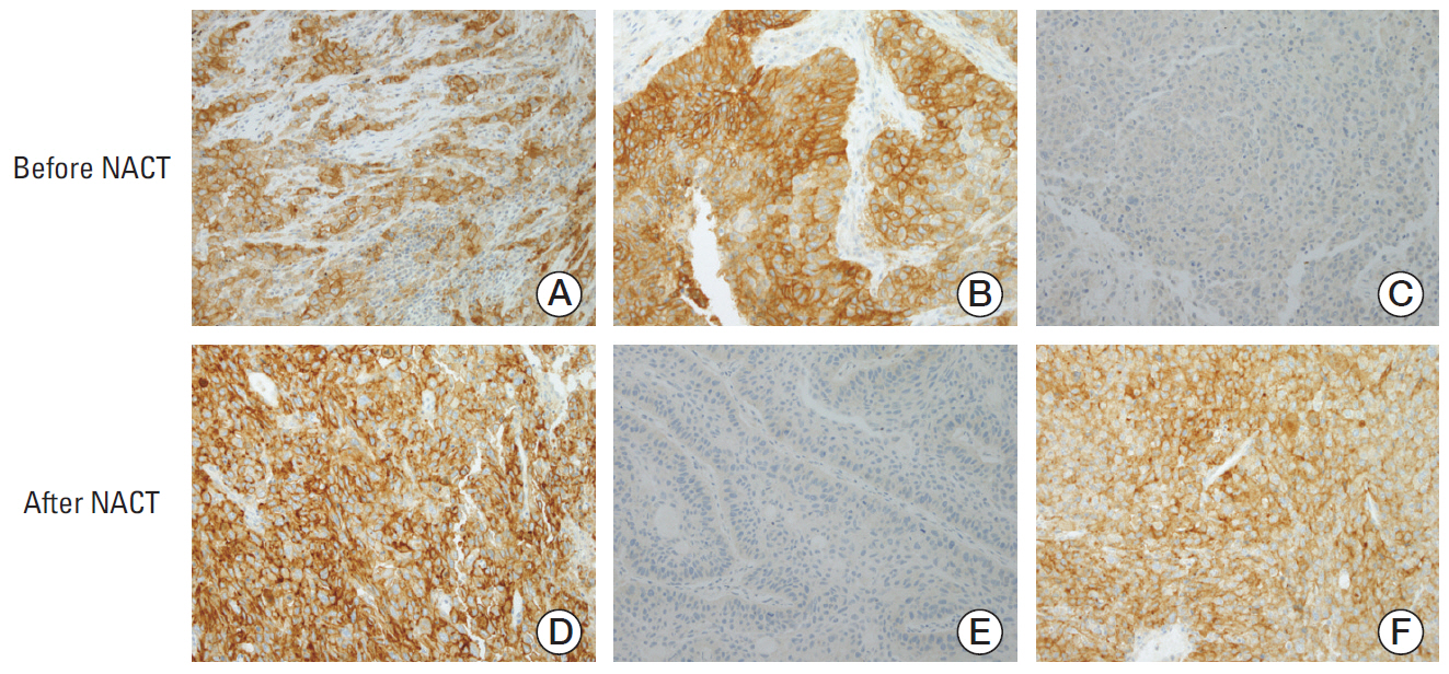
Before treatment, 41 (47.7%), 16 (18.6%), 19 (22.1%), and 10 (11.6%) patients were in the PD-L1 tumor proportion score ranges of < 1%, 1%-5%, 5%-50%, and ≥ 50%, respectively, in the landmark analysis cohort. The overall proportion of patients with positive tumor PD-L1 expression increased significantly after treatment with platinum-based NACT (45/86 [52.3%] before NACT vs. 65/86 [75.6%] after NACT; p=0.002) (Fig. 2A). That trend was consistent across subgroups stratified by sex, smoking history, histologic subtype, neoadjuvant treatment category, NACT regimen, and response to NACT (Fig. 2B-L), except for the subgroup of patients that achieved PR to NACT (Fig. 2K). The distributions of pre-NACT and post-NACT PD-L1 tumor proportion scores differed significantly within most subgroups, although they were only marginally different among males (Fig. 2B) and patients treated with taxane-platinum regimens (Fig. 2J). Notably, 26 patients were negative for tumor PD-L1 expression (tumor proportion score < 1%) before NACT and positive for tumor PD-L1 expression (tumor proportion score ≥ 1%) after NACT, whereas only six patients (7%) exhibited the reverse pattern (p < 0.001). Overall, 34 (39.5%), 12 (14%), and 40 (46.5%) patients showed an increased, decreased, or unchanged PD-L1 tumor proportion score following NACT, respectively (Fig. 3).
To assess the clinicopathologic characteristics associated with PD-L1 upregulation following NACT, we divided the patients into two groups based on the change in PD-L1 tumor proportion score following NACT. The ‘upward’ group included patients whose PD-L1 tumor proportion score increased after NACT. The ‘unchanged or downward’ group included the remaining patients. Comparison of the two groups indicated that the response to NACT was significantly correlated with the direction of change in PD-L1 tumor proportion score (p=0.015); patients who did not respond to NACT were twice as likely to show an upward change in PD-L1 tumor proportion score than those who achieved PR (Table 2). That result is in line with the results in Fig. 2K and L, which show that the proportion of tumor cells expressing PD-L1 increased after NACT only in tumors from patients who did not respond to NACT.
3. Prognostic impact of tumor PD-L1 upregulation following platinum-based NACT
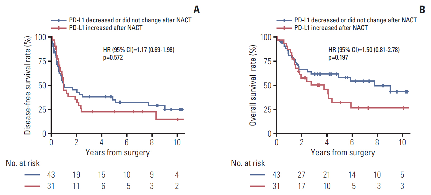
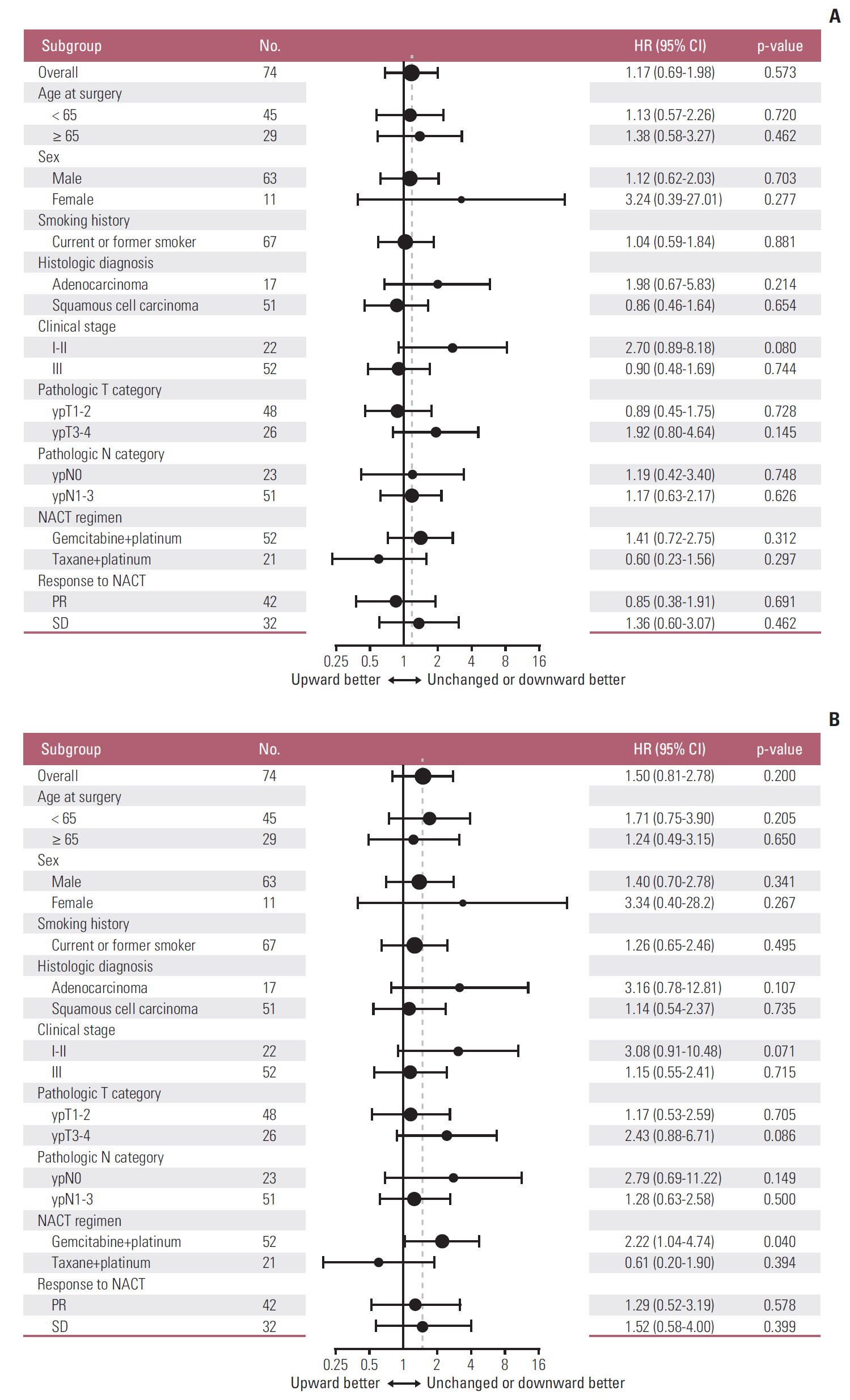
During a median follow-up of 7.3 years in the survival analysis cohort, 47 patients (63.5%) experienced disease recurrence and 41 patients (55.4%) died. DFS did not differ significantly between patients with increased PD-L1 tumor proportion score following NACT and those with unchanged or decreased PD-L1 tumor proportion score following NACT (median DFS, 11.8 months vs. 12 months) (Fig. 4A). Similarly, OS also did not differ significantly between the two groups (median OS, 3.8 years vs. 7.7 years) (Fig. 4B). Those results were consistent with multivariable Cox models adjusted for predefined sets of variables (Table 3). After adjustment, PD-L1 upregulation following NACT was not significantly associated with increased or decreased DFS and OS; however, visual inspection of the survival curves and the results of the multivariable Cox models indicated a trend toward poorer OS in patients whose PD-L1 tumor proportion score increased. That tendency was consistent across subgroups defined by sex, smoking history, histologic diagnosis, pretreatment clinical stage, pathologic T category assessed at surgery, NACT regimen, and response to NACT. The hazard ratio for death was less than one only in patients who received taxane-platinum combination therapy (Fig. 5).
We found that the PD-L1 tumor proportion score increased in a significant proportion of patients with NSCLC after treatment with platinum-based NACT. That pattern of change in tumor PD-L1 expression was consistent across clinically defined subgroups, except for the patients who achieved PR to NACT. Almost one-third of all the patients were negative for PD-L1 expression before NACT but positive for PD-L1 expression after NACT, based on a cutoff PD-L1 tumor proportion score of 1%, which is currently approved as an indication for pembrolizumab therapy after the failure of platinum-based doublet chemotherapy [3,14]. That result implies that the tumor PD-L1 expression level should be reassessed after platinum-based chemotherapy for appropriate selection of immunotherapy candidates. It also may support the design of clinical trials that incorporate PD-1/PD-L1 axis inhibitors into the treatment of patients with early-stage NSCLC.
A few retrospective studies recently investigated the alteration of tumor PD-L1 expression in patients with NSCLC who received platinum-based NACT (with or without radiotherapy) [9-13]. Those studies assessed much smaller numbers of paired tumor specimens than our study, or they focused only on a specific histologic subtype and therefore produced conflicting results. It is possible that tumor cells with PD-L1 downregulation as a result of exposure to platinum agents might undergo enhanced immune clearance, resulting in enrichment of PD-L1‒upregulated tumor cells in post-NACT tumor specimens. Indeed, our findings are consistent with the results of previous studies [8,11,13], although the small cohort size warrants confirmation in a larger cohort.
The predictive or prognostic value of tumor PD-L1 expression is an area of debate [24-28]. In our study, patients with increased PD-L1 tumor proportion score following platinum-based NACT had worse response to chemotherapy and tended to live for a shorter time than patients with unchanged or downregulated tumor PD-L1 expression, although the differences in prognosis were not statistically significant. Similarly, results from previous studies indicated that increased tumor PD-L1 expression following NACT was associated with poor response to chemotherapy and poor survival [9,10,13]. That, along with the frequent upregulation of tumor PD-L1 expression following platinum-based chemotherapy, suggests that patients with operable, early-stage or locally advanced NSCLC may benefit from postoperative PD-1/PD-L1 axis-inhibitor therapy after completion of standard neoadjuvant or adjuvant chemotherapy. The ongoing EORTC-ETOP phase 3 trial may confirm the validity of that hypothesis [29].
Our study has limitations. First and most importantly, the small numbers of patients included in the study cohorts limited the statistical power. Although we observed a consistent tendency toward shorter survival in patients with an upward change in tumor PD-L1 expression, the trend did not reach statistical significance. Additionally, the pre-NACT specimens were mostly obtained by percutaneous or transbronchial lung biopsy, whereas the post-NACT specimens were obtained by surgical resection. Therefore, there might be a systematic difference in measured tumor PD-L1 expression caused by a discrepancy in tissue acquisition techniques. Lastly, the study cohorts represented a heterogeneous patient population with various histologic subtypes and clinical stages. For that reason, most of the subgroups included only a small number of patients, which made it difficult to draw reliable conclusions from the subgroup analyses.
In summary, using matched pre-NACT and post-NACT tumor specimens obtained from patients with NSCLC, we demonstrated that the proportion of tumor cells expressing PD-L1 tended to increase after platinum-based chemotherapy, regardless of clinical characteristics and pathologic subtypes. The patients whose PD-L1 tumor proportion score increased following NACT tended to have poorer chemotherapy response and OS than patients whose PD-L1 tumor proportion score decreased or was unchanged. Our findings emphasize the need for reassessment of tumor PD-L1 expression after platinum-based chemotherapy and the potential benefit of postoperative PD-1/PD-L1 checkpoint inhibitor treatment for patients with operable NSCLC who undergo platinum-based neoadjuvant or adjuvant chemotherapy.

Conflict of interest relevant to this article was not reported.

Acknowledgements
This study was supported by the grants from the SNUBH Research Fund (grant number: 02-2014-042 to SHK), Korea Health Technology R&D Project through the Korea Health Industry Development Institute (KHIDI), funded by the Ministry of Health & Welfare, Republic of Korea (grant number: HI17C1290 to JHC) and the National Research Foundation of Korea (NRF) Grant funded by the Korean Government (MSIT) (No. 2017R1A5A1015626 to JHC). This manuscript has been edited by native English-speaking experts associated with BioScience Writers LLC, Houston, TX, USA.
Fig. 1.
Flow diagram of patient selection. NSCLC, non-small cell lung cancer; NACT, neoadjuvant chemotherapy; SNUBH, Seoul National University Bundang Hospital; IHC, immunohistochemistry; PD-L1, programmed death ligand 1.
crt-2018-537f1.jpg
Fig. 2.
Distribution of programmed death ligand 1 (PD-L1) tumor proportion score before and after neoadjuvant chemotherapy (NACT) in the landmark analysis cohort (A). Subgroup analyses of males (B), females (C), never-smokers (D), current or former smokers (E), adenocarcinoma (F), squamous cell carcinoma (G), patients who received NACT without radiotherapy (H), gemcitabine-platinum (I), taxane-platinum (J), patients with partial response to NACT (K), and patients with stable disease (L). Numbers in bar plots indicate the number of patients included in each PD-L1 tumor proportion score category.
crt-2018-537f2.jpg
Fig. 3.
Representative immunohistochemical staining patterns of programmed death ligand 1 (PD-L1) in tumor specimens. (A, D) PD-L1 tumor proportion score ≥ 50% before (A) and after (D) neoadjuvant chemotherapy (NACT). (B, E) PD-L1 tumor proportion score ≥ 50% before (B) and < 1% after (E) NACT. (C, F) PD-L1 tumor proportion score < 1% before (C) and ≥ 50% after (F) NACT (A-F, ×200).
crt-2018-537f3.jpg
Fig. 4.
Disease-free survival (A) and overall survival (B) according to the direction of change in programmed death ligand 1 (PD-L1) tumor proportion score following platinum-based neoadjuvant chemotherapy (NACT). HR, hazard ratio; CI, confidence interval.
crt-2018-537f4.jpg
Fig. 5.
Subgroup analyses of the prognostic value of increased (versus unchanged or decreased) programmed death ligand 1 tumor proportion score following platinum-based neoadjuvant chemotherapy (NACT) for disease-free survival (A) and overall survival (B). Note that subgroups with less than 10 patients are omitted. The vertical dashed line indicates the hazard ratio (HR) for all patients in the survival analysis cohort. CI, confidence interval; PR, partial response; SD, stable disease.
crt-2018-537f5.jpg
Table 1.
Patient characteristics
Characteristic Landmark analysis cohort (n=86) Survival analysis cohort (n=74)
Age at surgery (yr) 62 (37-79) 62 (37-79)
Sex
 Male 70 (81.4) 63 (85.1)
 Female 16 (18.6) 11 (14.9)
Smoking history
 Never-smoker 11 (12.8) 7 (9.5)
 Current or former smoker 75 (87.2) 67 (90.5)
Histologic diagnosis
 Adenocarcinoma 27 (31.4) 17 (23.0)
 Squamous cell carcinoma 53 (61.6) 51 (68.9)
 Othersa) 6 (7.0) 6 (8.1)
Pretreatment clinical stage
 I-II 22 (25.6) 22 (29.7)
 IIIA 40 (46.5) 40 (54.1)
 IIIB 12 (14.0) 12 (16.2)
 IV 12 (14.0)b) 0
Pathologic T category
 ypT1-2 57 (66.3) 48 (64.9)
 ypT3-4 29 (33.7) 26 (35.1)
Pathologic N category
 ypN0 24 (27.9) 23 (31.1)
 ypN1-3 62 (72.1) 51 (68.9)
Neoadjuvant treatment category
 NACT only 81 (94.2) 70 (94.6)
 Chemoradiation (sequential or concurrent) 5 (5.8) 4 (5.4)
NACT regimen
 Gemcitabine plus platinum 58 (67.4) 52 (70.3)
 Taxane plus platinum 24 (27.9) 21 (28.4)
 Othersc) 4 (4.7) 1 (1.4)
Best objective response to NACT
 PR 45 (52.3) 42 (56.8)
 SD 41 (47.7) 32 (43.2)

Values are presented as median (range) or number (%). NACT, neoadjuvant chemotherapy; PR, partial response; SD, stable disease.

a) Other histologic subtypes include large cell carcinoma (n=3), adenosquamous carcinoma (n=1), pleomorphic carcinoma (n=1), and sarcomatoid carcinoma (n=1),

b) Sites of distant metastases include brain (n=5), pleura (n=4), contralateral lung (n=2), and rib (n=1),

c) Other NACT regimens include pemetrexed plus platinum (n=2), etoposide plus platinum (n=1), and unknown regimen (n=1) in the landmark analysis cohort, and etoposide plus platinum (n=1) in the survival analysis cohort.

Table 2.
Direction of change in PD-L1 tumor proportion score following NACT in relation to clinicopathologic characteristics
Clinicopathologic variable Unchanged or downward (n=52) Upward (n=34) p-value
Age at surgery (yr) 62.5 (37-76) 61 (38-79) 0.572
Sex
 Male 46 (88.5) 24 (70.6) 0.049
 Female 6 (11.5) 10 (29.4)
Smoking history
 Never-smoker 5 (9.6) 6 (17.6) 0.331
 Current or former smoker 47 (90.4) 28 (82.4)
Histological diagnosis
 Adenocarcinoma 17 (32.7) 10 (29.4) 0.476
 Squamous cell carcinoma 30 (57.7) 23 (67.6)
 Others 5 (9.6) 1 (2.9)
Pretreatment clinical stage
 I-II 11 (21.2) 11 (32.4) 0.314
 III-IV 41 (78.8) 23 (67.6)
Pathologic T category
 ypT1-2 36 (69.2) 21 (61.8) 0.493
 ypT3-4 16 (30.8) 13 (38.2)
Pathologic N category
 ypN0 14 (26.9) 10 (29.4) 0.811
 ypN1-3 38 (73.1) 24 (70.6)
NACT regimen
 Gemcitabine plus platinum 36 (69.2) 22 (64.7) 0.644
 Taxane plus platinum 13 (25.0) 11 (32.4)
 Others 3 (5.8) 1 (2.9)
Best objective response to NACT
 PR 33 (63.5) 12 (35.3) 0.015
 SD 19 (36.5) 22 (64.7)

Values are presented as median (range) or number (%). PD-L1, programmed death ligand 1; NACT, neoadjuvant chemotherapy; PR, partial response; SD, stable disease.

Table 3.
Prognostic impact of PD-L1 upregulation (versus no change or downregulation) following platinum-based NACT
Model Disease-free survival
Overall survival
HR (95% CI) p-value HR (95% CI) p-value
Model 1a) 1.17 (0.67-2.03) 0.578 1.69 (0.89-3.22) 0.111
Model 2b) 1.32 (0.73-2.39) 0.366 1.60 (0.80-3.18) 0.182
Model 3c) 0.89 (0.46-1.70) 0.718 1.53 (0.69-3.38) 0.290

PD-L1, programmed death ligand 1; NACT, neoadjuvant chemotherapy; HR, hazard ratio; CI, confidence interval.

a) Model 1 is adjusted for age (≥ 65 vs. < 65), pretreatment clinical stage (I-II vs. III), and pathologic N category (ypN0 vs. ypN1 vs. ypN2 vs. ypN3),

b) Model 2 is adjusted for age (≥ 65 vs. < 65), pretreatment clinical stage (I-II vs. III), pathologic N category (ypN0 vs. ypN1 vs. ypN2 vs. ypN3), and pre-NACT PD-L1 tumor proportion score (< 1% vs. 1%-50% vs. ≥ 50%),

c) Model 3 is adjusted for age (≥ 65 vs. < 65), pretreatment clinical stage (I-II vs. III), pathologic N category (ypN0 vs. ypN1 vs. ypN2 vs. ypN3), and post-NACT PD-L1 tumor proportion score (< 1% vs. 1-50% vs. ≥ 50%).

  • 1. Reck M, Rodriguez-Abreu D, Robinson AG, Hui R, Csoszi T, Fulop A, et al. Pembrolizumab versus chemotherapy for PD-L1-positive non-small-cell lung cancer. N Engl J Med. 2016;375:1823–33. ArticlePubMed
  • 2. Gandhi L, Rodriguez-Abreu D, Gadgeel S, Esteban E, Felip E, De Angelis F, et al. Pembrolizumab plus chemotherapy in metastatic non-small-cell lung cancer. N Engl J Med. 2018;378:2078–92. ArticlePubMed
  • 3. Herbst RS, Baas P, Kim DW, Felip E, Perez-Gracia JL, Han JY, et al. Pembrolizumab versus docetaxel for previously treated, PD-L1-positive, advanced non-small-cell lung cancer (KEYNOTE-010): a randomised controlled trial. Lancet. 2016;387:1540–50. ArticlePubMed
  • 4. Brahmer J, Reckamp KL, Baas P, Crino L, Eberhardt WE, Poddubskaya E, et al. Nivolumab versus docetaxel in advanced squamous-cell non-small-cell lung cancer. N Engl J Med. 2015;373:123–35. ArticlePubMedPMC
  • 5. Borghaei H, Paz-Ares L, Horn L, Spigel DR, Steins M, Ready NE, et al. Nivolumab versus docetaxel in advanced nonsquamous non-small-cell lung cancer. N Engl J Med. 2015;373:1627–39. ArticlePubMedPMC
  • 6. Rittmeyer A, Barlesi F, Waterkamp D, Park K, Ciardiello F, von Pawel J, et al. Atezolizumab versus docetaxel in patients with previously treated non-small-cell lung cancer (OAK): a phase 3, open-label, multicentre randomised controlled trial. Lancet. 2017;389:255–65. ArticlePubMed
  • 7. Sul J, Blumenthal GM, Jiang X, He K, Keegan P, Pazdur R. FDA approval summary: pembrolizumab for the treatment of patients with metastatic non-small cell lung cancer whose tumors express programmed death-ligand 1. Oncologist. 2016;21:643–50. ArticlePubMedPMC
  • 8. Parra ER, Villalobos P, Behrens C, Jiang M, Pataer A, Swisher SG, et al. Effect of neoadjuvant chemotherapy on the immune microenvironment in non-small cell lung carcinomas as determined by multiplex immunofluorescence and image analysis approaches. J Immunother Cancer. 2018;6:48.ArticlePubMedPMCPDF
  • 9. Sheng J, Fang W, Yu J, Chen N, Zhan J, Ma Y, et al. Expression of programmed death ligand-1 on tumor cells varies pre and post chemotherapy in non-small cell lung cancer. Sci Rep. 2016;6:20090.ArticlePubMedPMCPDF
  • 10. Fujimoto D, Uehara K, Sato Y, Sakanoue I, Ito M, Teraoka S, et al. Alteration of PD-L1 expression and its prognostic impact after concurrent chemoradiation therapy in non-small cell lung cancer patients. Sci Rep. 2017;7:11373.ArticlePubMedPMCPDF
  • 11. Song Z, Yu X, Zhang Y. Altered expression of programmed death-ligand 1 after neo-adjuvant chemotherapy in patients with lung squamous cell carcinoma. Lung Cancer. 2016;99:166–71. ArticlePubMed
  • 12. Rojko L, Reiniger L, Teglasi V, Fabian K, Pipek O, Vagvolgyi A, et al. Chemotherapy treatment is associated with altered PD-L1 expression in lung cancer patients. J Cancer Res Clin Oncol. 2018;144:1219–26. ArticlePubMedPDF
  • 13. Zhang P, Ma Y, Lv C, Huang M, Li M, Dong B, et al. Upregulation of programmed cell death ligand 1 promotes resistance response in non-small-cell lung cancer patients treated with neo-adjuvant chemotherapy. Cancer Sci. 2016;107:1563–71. ArticlePubMedPMC
  • 14. Ettinger DS, Wood DE, Aisner DL, Akerley W, Bauman J, Chirieac LR, et al. Non-small cell lung cancer, version 5.2017, NCCN clinical practice guidelines in oncology. J Natl Compr Canc Netw. 2017;15:504–35. ArticlePubMed
  • 15. Kelland L. The resurgence of platinum-based cancer chemotherapy. Nat Rev Cancer. 2007;7:573–84. ArticlePubMedPDF
  • 16. Hato SV, Khong A, de Vries IJ, Lesterhuis WJ. Molecular pathways: the immunogenic effects of platinum-based chemotherapeutics. Clin Cancer Res. 2014;20:2831–7. ArticlePubMed
  • 17. Lesterhuis WJ, Punt CJ, Hato SV, Eleveld-Trancikova D, Jansen BJ, Nierkens S, et al. Platinum-based drugs disrupt STAT6-mediated suppression of immune responses against cancer in humans and mice. J Clin Invest. 2011;121:3100–8. ArticlePubMedPMC
  • 18. Rizvi NA, Hellmann MD, Brahmer JR, Juergens RA, Borghaei H, Gettinger S, et al. Nivolumab in combination with platinum-based doublet chemotherapy for first-line treatment of advanced non-small-cell lung cancer. J Clin Oncol. 2016;34:2969–79. ArticlePubMedPMC
  • 19. Liu SV, Camidge DR, Gettinger SN, Giaccone G, Heist RS, Hodi FS, et al. Long-term survival follow-up of atezolizumab in combination with platinum-based doublet chemotherapy in patients with advanced non-small-cell lung cancer. Eur J Cancer. 2018;101:114–22. ArticlePubMed
  • 20. Goldstraw P, Crowley J, Chansky K, Giroux DJ, Groome PA, Rami-Porta R, et al. The IASLC Lung Cancer Staging Project: proposals for the revision of the TNM stage groupings in the forthcoming (seventh) edition of the TNM Classification of malignant tumours. J Thorac Oncol. 2007;2:706–14. ArticlePubMed
  • 21. Herbst RS, Soria JC, Kowanetz M, Fine GD, Hamid O, Gordon MS, et al. Predictive correlates of response to the anti-PD-L1 antibody MPDL3280A in cancer patients. Nature. 2014;515:563–7. ArticlePubMedPMCPDF
  • 22. Fehrenbacher L, Spira A, Ballinger M, Kowanetz M, Vansteenkiste J, Mazieres J, et al. Atezolizumab versus docetaxel for patients with previously treated non-small-cell lung cancer (POPLAR): a multicentre, open-label, phase 2 randomised controlled trial. Lancet. 2016;387:1837–46. ArticlePubMed
  • 23. Eisenhauer EA, Therasse P, Bogaerts J, Schwartz LH, Sargent D, Ford R, et al. New response evaluation criteria in solid tumours: revised RECIST guideline (version 1.1). Eur J Cancer. 2009;45:228–47. ArticlePubMed
  • 24. Tsao MS, Le Teuff G, Shepherd FA, Landais C, Hainaut P, Filipits M, et al. PD-L1 protein expression assessed by immunohistochemistry is neither prognostic nor predictive of benefit from adjuvant chemotherapy in resected non-small cell lung cancer. Ann Oncol. 2017;28:882–9. ArticlePubMedPMCPDF
  • 25. Sun JM, Zhou W, Choi YL, Choi SJ, Kim SE, Wang Z, et al. Prognostic significance of PD-L1 in patients with non-small cell lung cancer: a large cohort study of surgically resected cases. J Thorac Oncol. 2016;11:1003–11. ArticlePubMed
  • 26. Wang A, Wang HY, Liu Y, Zhao MC, Zhang HJ, Lu ZY, et al. The prognostic value of PD-L1 expression for non-small cell lung cancer patients: a meta-analysis. Eur J Surg Oncol. 2015;41:450–6. ArticlePubMed
  • 27. Tokito T, Azuma K, Kawahara A, Ishii H, Yamada K, Matsuo N, et al. Predictive relevance of PD-L1 expression combined with CD8+ TIL density in stage III non-small cell lung cancer patients receiving concurrent chemoradiotherapy. Eur J Cancer. 2016;55:7–14. ArticlePubMed
  • 28. Cooper WA, Tran T, Vilain RE, Madore J, Selinger CI, Kohonen-Corish M, et al. PD-L1 expression is a favorable prognostic factor in early stage non-small cell carcinoma. Lung Cancer. 2015;89:181–8. ArticlePubMed
  • 29. O'Brien ME, Hasan B, Dafni U, Menis J, Peters S, De Waele M, et al. EORTC-ETOP randomized, phase 3 trial with anti-PD-1 monoclonal antibody pembrolizumab versus placebo for patients with early stage non-small cell lung cancer (NSCLC) after resection and standard adjuvant chemotherapy: PEARLS (NCT02504372). J Clin Oncol. 2016;34(15 Suppl):TPS8571.Article

Figure & Data

REFERENCES

    Citations

    Citations to this article as recorded by  
    • Prevalence and Clinical Association of CD276 (B7-H3) Expression in Pleural Mesothelioma: Results From the European Thoracic Platform Mesoscape Project
      Martina Haberecker, Jan H. Rüschoff, Charitini Andriakopoulou, Steven G. Gray, Kristiaan Nackaerts, Marc De Perrot, Luka Brcic, Ernest Nadal, Sotirios Tsimpoukis, Luca Ampollini, Joachim G. Aerts, Michaela B. Kirschner, Kim Monkhorst, Birgit Weynand, Fate
      JCO Precision Oncology.2025;[Epub]     CrossRef
    • Selenium-containing compounds, selenium nanoparticles and selenoproteins in the prevention and treatment of lung cancer
      Elena G. Varlamova
      Journal of Trace Elements in Medicine and Biology.2025; 88: 127620.     CrossRef
    • Potential Predictive Immune and Metabolic Biomarkers of Tumor Microenvironment Regarding Pathological and Clinical Response in Esophageal Cancer After Neoadjuvant Chemoradiotherapy: A Systematic Review
      H. H. Wang, E. N. Steffens, G. Kats-Ugurlu, B. van Etten, J. G. M. Burgerhof, G. A. P. Hospers, J. T. M. Plukker
      Annals of Surgical Oncology.2024; 31(1): 433.     CrossRef
    • Redetermination of PD-L1 expression after chemio-radiation in locally advanced PDL1 negative NSCLC patients: retrospective multicentric analysis
      Patrizia Ciammella, Salvatore Cozzi, Paolo Borghetti, Marco Galaverni, Valerio Nardone, Maria Paola Ruggieri, Matteo Sepulcri, Vieri Scotti, Alessio Bruni, Francesca Zanelli, Roberto Piro, Elena Tagliavini, Andrea Botti, Federico Iori, Emanuele Alì, Chiar
      Frontiers in Oncology.2024;[Epub]     CrossRef
    • First exploration of the on-treatment changes in tumor and organ uptake of a radiolabeled anti PD-L1 antibody during chemoradiotherapy in patients with non-small cell lung cancer using whole body PET
      Johanna E E Pouw, Sayed M S Hashemi, Marc C Huisman, Jessica E Wijngaarden, Maarten Slebe, Daniela E Oprea-Lager, Gerben J C Zwezerijnen, Danielle Vugts, Ezgi B Ulas, Tanja D de Gruijl, Teodora Radonic, Suresh Senan, C Willemien Menke-van der Houven van O
      Journal for ImmunoTherapy of Cancer.2024; 12(2): e007659.     CrossRef
    • Use of non-small cell lung cancer multicellular tumor spheroids to study the impact of chemotherapy
      Pauline Hulo, Sophie Deshayes, Judith Fresquet, Anne-Laure Chéné, Stéphanie Blandin, Nicolas Boisgerault, Jean-François Fonteneau, Lucas Treps, Marc G Denis, Jaafar Bennouna, Delphine Fradin, Elvire Pons-Tostivint, Christophe Blanquart
      Respiratory Research.2024;[Epub]     CrossRef
    • Alteration of PD-L1 (SP142) status after neoadjuvant chemotherapy and its clinical significance in triple-negative breast cancer
      Ji Won Woo, Eun Kyung Han, Koung Jin Suh, Se Hyun Kim, Jee Hyun Kim, So Yeon Park
      Breast Cancer Research and Treatment.2024; 207(2): 301.     CrossRef
    • Transforming Lung Cancer Management: A Promising Case Study of Immune Checkpoint Inhibitor Success Following a Multidisciplinary Approach
      Tadashi Nishimura, Hajime Fujimoto, Takumi Fujiwara, Tomohito Okano, Taro Yasuma, Esteban C. Gabazza, Hidenori Ibata, Tetsu Kobayashi
      Diagnostics.2024; 14(19): 2159.     CrossRef
    • Cryoablation plus chemotherapy regimen enhance anti-tumor immune response in a mouse model of Lewis lung cancer
      Hua Duan, Li Yang, Xueni Fang, Shaohua Yan, Yang Cao, Bingli Qiao, Tian Zhou, Kaiwen Hu
      International Journal of Hyperthermia.2024;[Epub]     CrossRef
    • Increased Tumor Mutation Burden Levels and Sensitivity of Non–Small Cell Lung Cancer to PD-L1 Blockade—Reply
      Biagio Ricciuti, Xinan Wang, Mark M. Awad
      JAMA Oncology.2023; 9(4): 570.     CrossRef
    • Add-On Effect of Hemagglutinating Virus of Japan Envelope Combined with Chemotherapy or Immune Checkpoint Inhibitor against Malignant Pleural Mesothelioma: An In Vivo Study
      Kazuma Sakura, Masao Sasai, Soichiro Funaki, Yasushi Shintani, Meinoshin Okumura, Yasufumi Kaneda
      Cancers.2023; 15(3): 929.     CrossRef
    • Predictive biomarkers for PD-1/PD-L1 checkpoint inhibitor response in NSCLC: an analysis of clinical trial and real-world data
      WeiQing Venus So, David Dejardin, Eva Rossmann, Jehad Charo
      Journal for ImmunoTherapy of Cancer.2023; 11(2): e006464.     CrossRef
    • Neoadjuvant treatment does not influence PD-L1 expression in stage III non-small-cell lung cancer: a retrospective analysis of tumor samples from the trials SAKK 16/96, 16/00, 16/01, and 16/14
      D. König, S. Savic Prince, S. Hayoz, P. Zens, S. Berezowska, W. Jochum, E. Stauffer, V. Braunersreuther, B. Trachsel, S. Thierstein, M. Mark, S. Schmid, A. Curioni-Fontecedro, A. Addeo, I. Opitz, M. Guckenberger, M. Früh, D.C. Betticher, H.-B. Ris, R. Stu
      ESMO Open.2023; 8(4): 101595.     CrossRef
    • Poliovirus receptor (PVR) mediates carboplatin-induced PD-L1 expression in non-small-cell lung cancer cells
      Chen Fu, Zongcai Liu, Taixue An, Haixia Li, Xiumei Hu, Xin Li, Xinyao Liu, Danjuan Wu, Ruyi Zhang, Kui Li, Yurong Qiu, Haifang Wang
      Biochimica et Biophysica Acta (BBA) - General Subjects.2023; 1867(10): 130439.     CrossRef
    • Remarkable Response to Immune Checkpoint Inhibitor Monotherapy in an EGFR-Mutant Pulmonary Adenocarcinoma Patient With 0% Expression of PD-L1
      Tetsuo Fujita, Hiroyuki Amano, Makoto Nakamura, Satoshi Hirano, Sukeyuki Nakamura
      Journal of Thoracic Oncology.2023; 18(9): e93.     CrossRef
    • Effect of Chemotherapeutics on In Vitro Immune Checkpoint Expression in Non-Small Cell Lung Cancer
      Yu Zhao, Zhe Wang, Xiuhuan Shi, Ting Liu, Wenwen Yu, Xiubao Ren, Hua Zhao
      Technology in Cancer Research & Treatment.2023;[Epub]     CrossRef
    • The Role of Regulatory T Cells in Cancer Treatment Resistance
      Anna Dąbrowska, Magdalena Grubba, Amar Balihodzic, Olga Szot, Bartosz Kamil Sobocki, Adrian Perdyan
      International Journal of Molecular Sciences.2023; 24(18): 14114.     CrossRef
    • Redirecting Chemotherapeutics to the Endoplasmic Reticulum Increases Tumor Immunogenicity and Potentiates Anti‐PD‐L1 Therapy
      Yucheng Xiang, Liqiang Chen, Chendong Liu, Xiaoli Yi, Lian Li, Yuan Huang
      Small.2022;[Epub]     CrossRef
    • FGFR3 Destabilizes PD-L1 via NEDD4 to Control T-cell–Mediated Bladder Cancer Immune Surveillance
      Weiqiang Jing, Ganyu Wang, Zhiwei Cui, Gaozhong Xiong, Xin Jiang, Yue Li, Wushan Li, Bo Han, Shouzhen Chen, Benkang Shi
      Cancer Research.2022; 82(1): 114.     CrossRef
    • The immune modifying effects of chemotherapy and advances in chemo-immunotherapy
      Daniel R. Principe, Suneel D. Kamath, Murray Korc, Hidayatullah G. Munshi
      Pharmacology & Therapeutics.2022; 236: 108111.     CrossRef
    • Adjuvant durvalumab for esophageal squamous cell carcinoma after neoadjuvant chemoradiotherapy: a placebo-controlled, randomized, double-blind, phase II study
      S. Park, J.-M. Sun, Y.-L. Choi, D. Oh, H.K. Kim, T. Lee, S.A. Chi, S.-H. Lee, Y.S. Choi, S.-H. Jung, M.-J. Ahn, Y.C. Ahn, K. Park, Y.M. Shim
      ESMO Open.2022; 7(1): 100385.     CrossRef
    • Analytical validation and initial clinical testing of quantitative microscopic evaluation for PD-L1 and HLA I expression on circulating tumor cells from patients with non-small cell lung cancer
      Jennifer L. Schehr, Nan Sethakorn, Zachery D. Schultz, Camila I. Hernandez, Rory M. Bade, Diego Eyzaguirre, Anupama Singh, David J. Niles, Leslie Henderson, Jay W. Warrick, Scott M. Berry, Kaitlin E. Sundling, David J. Beebe, Ticiana A. Leal, Joshua M. La
      Biomarker Research.2022;[Epub]     CrossRef
    • Translational Learnings in the Development of Chemo-Immunotherapy Combination to Bypass the Cold Tumor Microenvironment in Pancreatic Ductal Adenocarcinoma
      Hélène Kaplon
      Frontiers in Oncology.2022;[Epub]     CrossRef
    • Cost-effectiveness analyses of durvalumab consolidation therapy versus no consolidation therapy after chemoradiotherapy in stage-III NSCLC
      Salman Hussain, Jitka Klugarova, Miloslav Klugar
      Lung Cancer.2022; 170: 11.     CrossRef
    • Efficacy comparison of immune treating strategies for NSCLC patients with negative PD-L1 expression
      Kaiyue Ding, Minhan Yi, Hui Liang, Zhongkui Li, Yuan Zhang
      Expert Review of Clinical Immunology.2022; 18(7): 759.     CrossRef
    • Dual inhibition of TGFβ signaling and CSF1/CSF1R reprograms tumor-infiltrating macrophages and improves response to chemotherapy via suppressing PD-L1
      Tsung-Wei Chen, Wei-Ze Hung, Shu-Fen Chiang, William Tzu-Liang Chen, Tao-Wei Ke, Ji-An Liang, Chih-Yang Huang, Pei-Chen Yang, Kevin Chih-Yang Huang, K.S. Clifford Chao
      Cancer Letters.2022; 543: 215795.     CrossRef
    • SP142 PD-L1 Assays in Multiple Samples from the Same Patients with Early or Advanced Triple-Negative Breast Cancer
      Seung Ho Baek, Jee Hung Kim, Soong June Bae, Jung Hwan Ji, Yangkyu Lee, Joon Jeong, Yoon Jin Cha, Sung Gwe Ahn
      Cancers.2022; 14(13): 3042.     CrossRef
    • Case Report: Sustained complete remission on combination therapy with olaparib and pembrolizumab in BRCA2-mutated and PD-L1-positive metastatic cholangiocarcinoma after platinum derivate
      Taotao Zhou, Robert Mahn, Christian Möhring, Farsaneh Sadeghlar, Carsten Meyer, Marieta Toma, Barbara Kreppel, Markus Essler, Tim Glowka, Hanno Matthaei, Jörg C. Kalff, Christian P. Strassburg, Maria A. Gonzalez-Carmona
      Frontiers in Oncology.2022;[Epub]     CrossRef
    • Is there a place for chemotherapy in first line of treatment for metastatic NSCLC in era of immunotherapy?
      D. I. Yudin, K. K. Laktionov
      Medical alphabet.2022; (13): 18.     CrossRef
    • The effect of neoadjuvant therapy on PD-L1 expression and CD8+lymphocyte density in non-small cell lung cancer
      Philipp Zens, Corina Bello, Amina Scherz, Michael von Gunten, Adrian Ochsenbein, Ralph A. Schmid, Sabina Berezowska
      Modern Pathology.2022; 35(12): 1848.     CrossRef
    • MicroRNAs and Drug Resistance in Non-Small Cell Lung Cancer: Where Are We Now and Where Are We Going
      Roberto Cuttano, Miriam Kuku Afanga, Fabrizio Bianchi
      Cancers.2022; 14(23): 5731.     CrossRef
    • miRNome profiling of lung cancer metastases revealed a key role for miRNA-PD-L1 axis in the modulation of chemotherapy response
      Roberto Cuttano, Tommaso Colangelo, Juliana Guarize, Elisa Dama, Maria Pia Cocomazzi, Francesco Mazzarelli, Valentina Melocchi, Orazio Palumbo, Elena Marino, Elena Belloni, Francesca Montani, Manuela Vecchi, Massimo Barberis, Paolo Graziano, Andrea Pasqui
      Journal of Hematology & Oncology.2022;[Epub]     CrossRef
    • PD‐1 blockade using pembrolizumab in adolescent and young adult patients with advanced bone and soft tissue sarcoma
      Tahlia Scheinberg, Anna Lomax, Martin Tattersall, David Thomas, Geoff McCowage, Michael Sullivan, Rooshdiya Karim, Peter P. Luk, Annabelle Mahar, Fiona Bonar, Vivek A. Bhadri
      Cancer Reports.2021;[Epub]     CrossRef
    • PD‐L1 versus tumor mutation burden: Which is the better immunotherapy biomarker in advanced non‐small cell lung cancer?
      Aoran Dong, Yiming Zhao, Zhihua Li, Hai Hu
      The Journal of Gene Medicine.2021;[Epub]     CrossRef
    • Biomarkers predicting the response to chemotherapy and the prognosis in patients with esophageal squamous cell carcinoma
      Seiya Inoue, Takahiro Yoshida, Takeshi Nishino, Masakazu Goto, Mariko Aoyama, Naoya Kawakita, Yota Yamamoto, Furukita Yoshihito, Hiromitsu Takizawa, Akira Tangoku
      General Thoracic and Cardiovascular Surgery.2021; 69(3): 525.     CrossRef
    • Patient-Reported Outcomes with Durvalumab by PD-L1 Expression and Prior Chemoradiotherapy-Related Variables in Unresectable Stage III Non-Small-Cell Lung Cancer
      Marina C Garassino, Luis Paz-Ares, Rina Hui, Corinne Faivre-Finn, Alex Spira, David Planchard, Mustafa Özgüroğlu, Davey Daniel, David Vicente, Shuji Murakami, Corey Langer, Suresh Senan, David Spigel, Anna Rydén, Yiduo Zhang, Cathy O'Brien, Phillip A Denn
      Future Oncology.2021; 17(10): 1165.     CrossRef
    • Nivolumab with carboplatin, paclitaxel, and bevacizumab for first-line treatment of advanced nonsquamous non-small-cell lung cancer
      S. Sugawara, J.-S. Lee, J.-H. Kang, H.R. Kim, N. Inui, T. Hida, K.H. Lee, T. Yoshida, H. Tanaka, C.-T. Yang, M. Nishio, Y. Ohe, T. Tamura, N. Yamamoto, C.-J. Yu, H. Akamatsu, Y. Namba, N. Sumiyoshi, K. Nakagawa
      Annals of Oncology.2021; 32(9): 1137.     CrossRef
    • Temporal evolution of programmed death-ligand 1 expression in patients with non-small cell lung cancer
      Chang Hyun Nam, Jaemoon Koh, Chan-Young Ock, Miso Kim, Bhumsuk Keam, Tae Min Kim, Yoon Kyung Jeon, Dong-Wan Kim, Doo Hyun Chung, Dae Seog Heo
      The Korean Journal of Internal Medicine.2021; 36(4): 975.     CrossRef
    • Methylseleninic acid overcomes programmed death‐ligand 1‐mediated resistance of prostate cancer and lung cancer
      Wenli Hu, Yurong Ma, Chong Zhao, Shutao Yin, Hongbo Hu
      Molecular Carcinogenesis.2021; 60(11): 746.     CrossRef
    • Immunological Status of Bladder Cancer Patients Based on Urine Leukocyte Composition at Radical Cystectomy
      Elisabet Cantó, Óscar Rodríguez Faba, Carlos Zamora, Maria Mulet, Maria Soledad Garcia-Cuerva, Ana Palomino, Georgia Anguera, Alberto Breda, Pablo Maroto, Sílvia Vidal
      Biomedicines.2021; 9(9): 1125.     CrossRef
    • Ex vivo expanded allogeneic natural killer cells have potent cytolytic activity against cancer cells through different receptor-ligand interactions
      Daun Jung, Young Seok Baek, In Jee Lee, Ki Yeon Kim, Heejoo Jang, Sohyun Hwang, Jieun Jung, Yong-wha Moon, Kyung-Soon Park, Yong-Soo Choi, Hee Jung An
      Journal of Experimental & Clinical Cancer Research.2021;[Epub]     CrossRef
    • Prognostic significance of T‐cell–inflamed gene expression profile and PD‐L1 expression in patients with esophageal cancer
      Torben Steiniche, Sun Young Rha, Hyun Cheol Chung, Jeanette Baehr Georgsen, Morten Ladekarl, Marianne Nordsmark, Marie Louise Jespersen, Hyo Song Kim, Hyunki Kim, Carly Fein, Laura H. Tang, Ting Wu, Matthew J. Marton, Senaka Peter, David P. Kelsen, Geoffr
      Cancer Medicine.2021; 10(23): 8365.     CrossRef
    • A novel form of immunotherapy using antigen peptides conjugated on PD-L1 antibody
      Eun Ji Lee, Gun-Young Jang, Sung Eun Lee, Ji won Lee, Hee Dong Han, Yeong-Min Park, Tae Heung Kang
      Immunology Letters.2021;[Epub]     CrossRef
    • Assessment of the expression of the immune checkpoint molecules PD‐1, CTLA4, TIM‐3 and LAG‐3 across different cancers in relation to treatment response, tumor‐infiltrating immune cells and survival
      Lei Tu, Renguo Guan, Hanting Yang, Yu Zhou, Weifeng Hong, Liheng Ma, Guangzhe Zhao, Min Yu
      International Journal of Cancer.2020; 147(2): 423.     CrossRef
    • Smokers or non-smokers: who benefits more from immune checkpoint inhibitors in treatment of malignancies? An up-to-date meta-analysis
      Jiahang Mo, Xiao Hu, Lihu Gu, Bangsheng Chen, Parikshit Asutosh Khadaroo, Zefeng Shen, Lei Dong, Yuqi Lv, Marylin Nyaradzo Chitumba, Jiequan Liu
      World Journal of Surgical Oncology.2020;[Epub]     CrossRef
    • Prognostic value of PD-L1 expression on tumor cells combined with CD8+ TIL density in patients with locally advanced non-small cell lung cancer treated with concurrent chemoradiotherapy
      Kathrin Gennen, Lukas Käsmann, Julian Taugner, Chukwuka Eze, Monika Karin, Olarn Roengvoraphoj, Jens Neumann, Amanda Tufman, Michael Orth, Simone Reu, Claus Belka, Farkhad Manapov
      Radiation Oncology.2020;[Epub]     CrossRef
    • PD-L1 induction in tumor tissue after hypofractionated thoracic radiotherapy for non-small cell lung cancer
      Jaanika Narits, Hannes Tamm, Jana Jaal
      Clinical and Translational Radiation Oncology.2020; 22: 83.     CrossRef
    • Variation of PD-L1 expression in locally advanced cervical cancer following neoadjuvant chemotherapy
      Yun Liang, Minghua Yu, Caiyun Zhou, Xiaojun Zhu
      Diagnostic Pathology.2020;[Epub]     CrossRef
    • Application of immune checkpoint inhibitors in EGFR-mutant non-small-cell lung cancer: from bed to bench
      Rui Jin, Jing Zhao, Lexin Xia, Qin Li, Wen Li, Ling Peng, Yang Xia
      Therapeutic Advances in Medical Oncology.2020;[Epub]     CrossRef
    • Independent Prognostic Value of Intratumoral Heterogeneity and Immune Response Features by Automated Digital Immunohistochemistry Analysis in Early Hormone Receptor-Positive Breast Carcinoma
      Dovile Zilenaite, Allan Rasmusson, Renaldas Augulis, Justinas Besusparis, Aida Laurinaviciene, Benoit Plancoulaine, Valerijus Ostapenko, Arvydas Laurinavicius
      Frontiers in Oncology.2020;[Epub]     CrossRef
    • Immunostimulation with chemotherapy in the era of immune checkpoint inhibitors
      Lorenzo Galluzzi, Juliette Humeau, Aitziber Buqué, Laurence Zitvogel, Guido Kroemer
      Nature Reviews Clinical Oncology.2020; 17(12): 725.     CrossRef
    • Re-biopsy after first line treatment in advanced NSCLC can reveal changes in PD-L1 expression
      Malene Støchkel Frank, Uffe Bødtger, Asbjørn Høegholm, Inger Merete Stamp, Julie Gehl
      Lung Cancer.2020; 149: 23.     CrossRef
    • The Mechanisms of PD-L1 Regulation in Non-Small-Cell Lung Cancer (NSCLC): Which Are the Involved Players?
      Giuseppe Lamberti, Monia Sisi, Elisa Andrini, Arianna Palladini, Francesca Giunchi, Pier-Luigi Lollini, Andrea Ardizzoni, Francesco Gelsomino
      Cancers.2020; 12(11): 3129.     CrossRef
    • The Dichotomous Role of Bone Marrow Derived Cells in the Chemotherapy-Treated Tumor Microenvironment
      Avital Vorontsova, Tal Kan, Ziv Raviv, Yuval Shaked
      Journal of Clinical Medicine.2020; 9(12): 3912.     CrossRef
    • Adjuvant Chemotherapy Increases Programmed Death-Ligand 1 (PD-L1) Expression in Non–small Cell Lung Cancer Recurrence
      Max Lacour, Stefanie Hiltbrunner, Seok-Yun Lee, Alex Soltermann, Elisabeth Jane Rushing, Davide Soldini, Walter Weder, Alessandra Curioni-Fontecedro
      Clinical Lung Cancer.2019; 20(5): 391.     CrossRef
    • PD-L1 Testing in Non-small Cell Lung Cancer: Past, Present, and Future
      Hyojin Kim, Jin-Haeng Chung
      Journal of Pathology and Translational Medicine.2019; 53(4): 199.     CrossRef

    • PubReader PubReader
    • ePub LinkePub Link
    • Cite
      CITE
      export Copy Download
      Close
      Download Citation
      Download a citation file in RIS format that can be imported by all major citation management software, including EndNote, ProCite, RefWorks, and Reference Manager.

      Format:
      • RIS — For EndNote, ProCite, RefWorks, and most other reference management software
      • BibTeX — For JabRef, BibDesk, and other BibTeX-specific software
      Include:
      • Citation for the content below
      Effect of Platinum-Based Chemotherapy on PD-L1 Expression on Tumor Cells in Non-small Cell Lung Cancer
      Cancer Res Treat. 2019;51(3):1086-1097.   Published online November 5, 2018
      Close
    • XML DownloadXML Download
    Effect of Platinum-Based Chemotherapy on PD-L1 Expression on Tumor Cells in Non-small Cell Lung Cancer
    Image Image Image Image Image
    Fig. 1. Flow diagram of patient selection. NSCLC, non-small cell lung cancer; NACT, neoadjuvant chemotherapy; SNUBH, Seoul National University Bundang Hospital; IHC, immunohistochemistry; PD-L1, programmed death ligand 1.
    Fig. 2. Distribution of programmed death ligand 1 (PD-L1) tumor proportion score before and after neoadjuvant chemotherapy (NACT) in the landmark analysis cohort (A). Subgroup analyses of males (B), females (C), never-smokers (D), current or former smokers (E), adenocarcinoma (F), squamous cell carcinoma (G), patients who received NACT without radiotherapy (H), gemcitabine-platinum (I), taxane-platinum (J), patients with partial response to NACT (K), and patients with stable disease (L). Numbers in bar plots indicate the number of patients included in each PD-L1 tumor proportion score category.
    Fig. 3. Representative immunohistochemical staining patterns of programmed death ligand 1 (PD-L1) in tumor specimens. (A, D) PD-L1 tumor proportion score ≥ 50% before (A) and after (D) neoadjuvant chemotherapy (NACT). (B, E) PD-L1 tumor proportion score ≥ 50% before (B) and < 1% after (E) NACT. (C, F) PD-L1 tumor proportion score < 1% before (C) and ≥ 50% after (F) NACT (A-F, ×200).
    Fig. 4. Disease-free survival (A) and overall survival (B) according to the direction of change in programmed death ligand 1 (PD-L1) tumor proportion score following platinum-based neoadjuvant chemotherapy (NACT). HR, hazard ratio; CI, confidence interval.
    Fig. 5. Subgroup analyses of the prognostic value of increased (versus unchanged or decreased) programmed death ligand 1 tumor proportion score following platinum-based neoadjuvant chemotherapy (NACT) for disease-free survival (A) and overall survival (B). Note that subgroups with less than 10 patients are omitted. The vertical dashed line indicates the hazard ratio (HR) for all patients in the survival analysis cohort. CI, confidence interval; PR, partial response; SD, stable disease.
    Effect of Platinum-Based Chemotherapy on PD-L1 Expression on Tumor Cells in Non-small Cell Lung Cancer
    Characteristic Landmark analysis cohort (n=86) Survival analysis cohort (n=74)
    Age at surgery (yr) 62 (37-79) 62 (37-79)
    Sex
     Male 70 (81.4) 63 (85.1)
     Female 16 (18.6) 11 (14.9)
    Smoking history
     Never-smoker 11 (12.8) 7 (9.5)
     Current or former smoker 75 (87.2) 67 (90.5)
    Histologic diagnosis
     Adenocarcinoma 27 (31.4) 17 (23.0)
     Squamous cell carcinoma 53 (61.6) 51 (68.9)
     Othersa) 6 (7.0) 6 (8.1)
    Pretreatment clinical stage
     I-II 22 (25.6) 22 (29.7)
     IIIA 40 (46.5) 40 (54.1)
     IIIB 12 (14.0) 12 (16.2)
     IV 12 (14.0)b) 0
    Pathologic T category
     ypT1-2 57 (66.3) 48 (64.9)
     ypT3-4 29 (33.7) 26 (35.1)
    Pathologic N category
     ypN0 24 (27.9) 23 (31.1)
     ypN1-3 62 (72.1) 51 (68.9)
    Neoadjuvant treatment category
     NACT only 81 (94.2) 70 (94.6)
     Chemoradiation (sequential or concurrent) 5 (5.8) 4 (5.4)
    NACT regimen
     Gemcitabine plus platinum 58 (67.4) 52 (70.3)
     Taxane plus platinum 24 (27.9) 21 (28.4)
     Othersc) 4 (4.7) 1 (1.4)
    Best objective response to NACT
     PR 45 (52.3) 42 (56.8)
     SD 41 (47.7) 32 (43.2)
    Clinicopathologic variable Unchanged or downward (n=52) Upward (n=34) p-value
    Age at surgery (yr) 62.5 (37-76) 61 (38-79) 0.572
    Sex
     Male 46 (88.5) 24 (70.6) 0.049
     Female 6 (11.5) 10 (29.4)
    Smoking history
     Never-smoker 5 (9.6) 6 (17.6) 0.331
     Current or former smoker 47 (90.4) 28 (82.4)
    Histological diagnosis
     Adenocarcinoma 17 (32.7) 10 (29.4) 0.476
     Squamous cell carcinoma 30 (57.7) 23 (67.6)
     Others 5 (9.6) 1 (2.9)
    Pretreatment clinical stage
     I-II 11 (21.2) 11 (32.4) 0.314
     III-IV 41 (78.8) 23 (67.6)
    Pathologic T category
     ypT1-2 36 (69.2) 21 (61.8) 0.493
     ypT3-4 16 (30.8) 13 (38.2)
    Pathologic N category
     ypN0 14 (26.9) 10 (29.4) 0.811
     ypN1-3 38 (73.1) 24 (70.6)
    NACT regimen
     Gemcitabine plus platinum 36 (69.2) 22 (64.7) 0.644
     Taxane plus platinum 13 (25.0) 11 (32.4)
     Others 3 (5.8) 1 (2.9)
    Best objective response to NACT
     PR 33 (63.5) 12 (35.3) 0.015
     SD 19 (36.5) 22 (64.7)
    Model Disease-free survival
    Overall survival
    HR (95% CI) p-value HR (95% CI) p-value
    Model 1a) 1.17 (0.67-2.03) 0.578 1.69 (0.89-3.22) 0.111
    Model 2b) 1.32 (0.73-2.39) 0.366 1.60 (0.80-3.18) 0.182
    Model 3c) 0.89 (0.46-1.70) 0.718 1.53 (0.69-3.38) 0.290
    Table 1. Patient characteristics

    Values are presented as median (range) or number (%). NACT, neoadjuvant chemotherapy; PR, partial response; SD, stable disease.

    Other histologic subtypes include large cell carcinoma (n=3), adenosquamous carcinoma (n=1), pleomorphic carcinoma (n=1), and sarcomatoid carcinoma (n=1),

    Sites of distant metastases include brain (n=5), pleura (n=4), contralateral lung (n=2), and rib (n=1),

    Other NACT regimens include pemetrexed plus platinum (n=2), etoposide plus platinum (n=1), and unknown regimen (n=1) in the landmark analysis cohort, and etoposide plus platinum (n=1) in the survival analysis cohort.

    Table 2. Direction of change in PD-L1 tumor proportion score following NACT in relation to clinicopathologic characteristics

    Values are presented as median (range) or number (%). PD-L1, programmed death ligand 1; NACT, neoadjuvant chemotherapy; PR, partial response; SD, stable disease.

    Table 3. Prognostic impact of PD-L1 upregulation (versus no change or downregulation) following platinum-based NACT

    PD-L1, programmed death ligand 1; NACT, neoadjuvant chemotherapy; HR, hazard ratio; CI, confidence interval.

    Model 1 is adjusted for age (≥ 65 vs. < 65), pretreatment clinical stage (I-II vs. III), and pathologic N category (ypN0 vs. ypN1 vs. ypN2 vs. ypN3),

    Model 2 is adjusted for age (≥ 65 vs. < 65), pretreatment clinical stage (I-II vs. III), pathologic N category (ypN0 vs. ypN1 vs. ypN2 vs. ypN3), and pre-NACT PD-L1 tumor proportion score (< 1% vs. 1%-50% vs. ≥ 50%),

    Model 3 is adjusted for age (≥ 65 vs. < 65), pretreatment clinical stage (I-II vs. III), pathologic N category (ypN0 vs. ypN1 vs. ypN2 vs. ypN3), and post-NACT PD-L1 tumor proportion score (< 1% vs. 1-50% vs. ≥ 50%).


    Cancer Res Treat : Cancer Research and Treatment
    Close layer
    TOP