Skip Navigation
Skip to contents

Cancer Res Treat : Cancer Research and Treatment

OPEN ACCESS

Articles

Page Path
HOME > Cancer Res Treat > Volume 51(2); 2019 > Article
Original Article Prognostic Role and Clinical Association of Tumor-Infiltrating Lymphocyte, Programmed Death Ligand-1 Expression with Neutrophil-Lymphocyte Ratio in Locally Advanced Triple-Negative Breast Cancer
Jieun Lee, MD, PhD1,2, Dong-Min Kim, BS3, Ahwon Lee, MD, PhD2,3,
Cancer Research and Treatment : Official Journal of Korean Cancer Association 2019;51(2):649-663.
DOI: https://doi.org/10.4143/crt.2018.270
Published online: July 31, 2018

1Division of Medical Oncology, Department of Internal Medicine, College of Medicine, The Catholic University of Korea, Seoul, Korea

2Cancer Research Institute, The Catholic University of Korea, Seoul, Korea

3Department of Hospital Pathology, College of Medicine, The Catholic University of Korea, Seoul, Korea

Correspondence: Ahwon Lee, MD, PhD Department of Hospital Pathology, College of Medicine, The Catholic University of Korea, 222 Banpo-daero, Seocho-gu, Seoul 06591, Korea
Tel: 82-2-2258-1621 Fax: 82-2-2258-1627 E-mail: klee@catholic.ac.kr
• Received: May 10, 2018   • Accepted: July 26, 2018

Copyright © 2019 by the Korean Cancer Association

This is an Open Access article distributed under the terms of the Creative Commons Attribution Non-Commercial License (http://creativecommons.org/licenses/by-nc/4.0/) which permits unrestricted non-commercial use, distribution, and reproduction in any medium, provided the original work is properly cited.

  • 11,991 Views
  • 577 Download
  • 56 Web of Science
  • 46 Crossref
  • 60 Scopus
prev next
  • Purpose
    Tumor-infiltrating lymphocyte (TIL), programmed death-ligand 1 (PD-L1) expression and neutrophil-to-lymphocyte ratio (NLR) is associated to immunogenicity and prognosis of breast cancer. We analyzed baseline NLR, changes of NLR, TIL, and PD-L1 during neoadjuvant chemotherapy (NAC) and their clinical implication in triple-negative breast cancer (TNBC).
  • Materials and Methods
    Between January 2008 to December 2015, 358 TNBC patients were analyzed. Baseline NLR, 50 paired NLR (initial diagnosis, after completion of NAC) and 34 paired tissues (initial diagnosis, surgical specimen after completion of NAC) were collected. Changes of TIL, CD4, CD8, forkhead box P3 (FOXP3), and PD-L1 expression were assessed with immunohistochemical stain.
  • Results
    Low NLR (≤ 3.16) was associated to superior survival (overall survival: 41.83 months vs. 36.5 months, p=0.002; disease-free survival [DFS]: 37.85 months vs. 32.14 months, p=0.032). Modest NLR change after NAC (–30% < NLR change < 100%) showed prolonged DFS (38.37 months vs. 22.37 months, p=0.015). During NAC, negative or negative conversion of tumor PD-L1 expression was associated to poor DFS (34.77 months vs. 16.03 months, p=0.037), and same or increased TIL showed trends for superior DFS, but without statistical significance. Positive tumor PD-L1 expression (H-score ≥ 5) in baseline or post-NAC tissue was associated to superior DFS (57.6 months vs. 12.5 months, p=0.001 and 53.3 months vs. 18.9 months, p=0.040). Positive stromal PD-L1 expression in baseline was also associated to superior DFS (50.2 months vs. 20.4 months, p=0.002).
  • Conclusion
    In locally advanced TNBC, baseline NLR, changes of NLR during NAC was associated to survival. Baseline PD-L1 expression and changes of PD-L1 expression in tumor tissue during NAC also showed association to prognosis.
Triple-negative breast cancer (TNBC) accounts for 10 to 20% of total breast cancer and is defined as absence of estrogen receptor (ER), progesterone receptor (PR), and human epidermal growth factor-2 (HER-2) expression by immunohistochemical stain. Compared to other subtypes, TNBC shows relatively poorer outcome due to its aggressive clinical behavior and lack of optimal molecular target for effective treatment [1]. Molecular profiling may more accurately predict the prognosis and response to chemotherapy of TNBC [2], but genomic profiling is not routinely feasible in clinical setting [3]. Other than genomic analysis, various clinical, laboratory and pathologic parameters are assessed for convenient prediction of chemotherapy response and overall prognosis of TNBC.
Inflammation is traditionally known to be associated with oncogenesis and progression in various malignancies. Systemic inflammation and local immune response both reflect prognosis of many solid cancers, and cross-talk between two immune response is suspected in progression of malignancies [4]. Neutrophil-lymphocyte ratio (NLR) represents systemic inflammation, and there are various reports supporting NLR reflects prognosis in solid cancers [4], including breast cancer [3,5,6]. In locally advanced breast cancer, pretreatment NLR is reported to be associated with superior response to neoadjuvant chemotherapy (NAC) and disease-free survival (DFS) [3,7]. Tumor-infiltrating lymphocyte (TIL) is suggested to represent anti-tumor immune response in tumor environment and associated with prognosis in various cancers including breast cancer [8-10]. In breast cancer patients who receive NAC, the presence of TIL and the changes of T cell marker is reported to be associated to patients’ prognosis [11]. Programmed cell death receptor 1 (PD-1), programmed cell death receptor ligand-1 (PD-L1) is one of a major molecule regulating T-cell activation and immune surveillance [12]. PD-L1 is expressed in both tumor cell and stroma, with heterogeneous distribution between cancers, including breast cancer [13]. PD-L1 expression is reported to be correlated to TIL and response to NAC in breast cancer [14] and other literatures report that expression of PD-L1 may be associated to prognosis in breast cancer, but with conflicting results [15,16].
The association of NLR, TIL, or PD-L1 with clinicopathologic parameters or survival outcomes were previously studied in various cancers [5-7,9,10,17]. NLR, TIL, and PD-L1 expression in breast cancer each has been studied as predictive or prognostic marker for estimating the response of NAC, DFS, or overall survival (OS) in various literatures [3,7,14,17]. Among NLR, TIL, and PD-L1, the role or NLR was most extensively studied in breast cancer, but the patient population was heterogeneous with mixture of subtypes or commonly studied in hormone receptor positive breast cancer [3,6,7]. The expression or changes of TIL during chemotherapy is traditionally known to be associated to tumor response in breast cancer [18], and the association between TIL and PD-L1 expression among tumor tissues are recently updated, but with heterogeneous reports and analyses methods [14,17,19,20]. Additionally, the role of PD-L1 in predicting prognosis and tumor response in breast canceris reported with conflicting results [20,21]. The interaction between systemic inflammatory marker such as NLR and local inflammatory marker such as TIL or PD-L1 has not been studied in-depth previously. Immunogenicity and related tumor response and prognosis is becoming more important in breast cancer, and administration of NAC may potentiate the changes of immunogenicity in breast cancer population. Therefore, we investigated the role of NLR, TIL, and PD-1 in homogeneous TNBC patient population.
In this study, we primarily analyzed the association of NLR with survival outcomes in early or locally advanced TNBC. Furthermore, the association between clinical outcomes, TIL and PD-L1 was analyzed in subgroup of patients who received NAC. Interrelationship between NLR, TIL, CD4+ TIL, CD8+ TIL, forkhead box P3 (FOXP3)+ TIL, and PD-L1 expression was also studied among NAC patients.
1. Patients
From January 2007 to December 2015, the medical records of 358 patients who were diagnosed as TNBC in Seoul St. Mary’s Hospital were retrospectively reviewed. All patients were pathologically diagnosed as stage I to III and received adequate local, systemic treatment. Triple negative breast cancer was defined as ER, PR, and HER2 all negative by American Society of Clinical. Oncology–College of American Pathologists guideline [22,23]; ER and PR negative defined as < 1% tumor cells displaying nuclear staining. HER2 negativity defined as HER2 immunohistochemistry 0/1 (no staining, faint/barely perceptible membrane staining, or weak incomplete membrane staining in < 10% of invasive tumor cells) or HER2 silver in situ hybridization negative (Dual-probe HER2/Chr17 ratio < 2.0 with an average HER2 copy number < 4.0 signals/cell). Among 358 patients, 50 patients received NAC. Baseline complete blood count (CBC) was collected before surgical treatment. The other eligible criteria were as follows: (1) adequate bone marrow function without hematologic disease; (2) patients who regularly followed up in Seoul St. Mary’s Hospital. In 50 patients who received NAC, baseline CBC was collected at initial pathologic diagnosis, and CBC was recollected before patient went through surgical treatment after completion of NAC. Among 34 patients who received NAC, baseline CBC, pre-operative CBC, biopsy specimen obtained during initial diagnosis and surgical pathologic specimens were collected.
2. Treatments
Initial pathologic diagnosis, laboratory evaluation and staging were done within 2 weeks since initial patient visit. In patient population who received primary breast conserving surgery or total mastectomy with axillary node staging, they received adequate anthracycline or taxane based adjuvant chemotherapy or standard CMF regimen and radiation therapy. Adjuvant chemotherapy regimens are as follows: (1) adriamycin (60 mg/m2 intravenously, day 1) and cyclophosphamide (600 mg/m2 intravenously, day 1) every 3 weeks for 4 cycles followed by docetaxel (75 mg/m2 intravenously, day 1) every 3 weeks for 4 cycles (AC-T); (2) adriamycin (60 mg/m2 intravenously, day 1) and cyclophosphamide (600 mg/m2 intravenously, day 1) (AC) every 3 weeks for 4 cycles; (3) docetaxel (75 mg/m2 intravenously, day 1) and adriamycin (50 mg/m2 intravenously, day 1) (DA) every 3 weeks for 2-4 cycles; (4) docetaxel (75 mg/m2 intravenously, day 1) and cyclophosphamide (600 mg/m2 intravenously, day 1) (TC) every 3 weeks for 4 cycles; (5) docetaxel (75 mg/m2 intravenously, day 1), adriamycin (50 mg/m2 intravenously, day 1), and cyclophosphamide (500 mg/m2 intravenously, day 1) (TAC) every 3 weeks for 6 cycles; (6) 5-fluorouracil (5-FU; 600 mg/m2 intravenously, day 1), epirubiclin (60 mg/m2 intravenously, day 1), and cyclophosphamide (600 mg/m2 intravenously, day 1) (FEC); (7) oral cyclophosphamide (100 mg/m2 intravenously, day 1 to 14), methotrexate (40 mg/m2 intravenously, day 1 and 8), and 5-FU (600 mg/m2 IV, day 1 and 8) (CMF) every 4 weeks for 6 cycles. Among patients who were diagnosed as stage II or III breast cancer, part of patients received anthracycline and taxane based NAC followed by surgery and adjuvant radiation. Anthracycline and taxane based NAC regimen is as follows: (1) docetaxel (75 mg/m2 intravenously, day 1) and adriamycin (50 mg/m2 intravenously, day 1) (DA) every 3 weeks for 2-6 cycles; (2) adriamycin (60 mg/m2 intravenously, day 1) and cyclophosphamide (600 mg/m2 intravenously, day 1) (AC) every 3 weeks for 4 cycles; (3) epirubicin (75 mg/m2 intravenously, day 1) and cyclophosphamide (600 mg/m2 intravenously, day 1) (EC) for 4 cycles. Patients were followed by every 3 months with history taking and physical examination. Laboratory evaluation including serum cancer antigen 15-3 and carcinoembryonic antigen, chest and abdominal computed tomography (CT) scan were done if clinically warranted. Mammography and breast sonography was done every six months for follow-up (S1 Fig.).
3. Definition of neutrophil-to-lymphocyte ratio
Neutrophil-to-lymphocyte (NLR) ratio was determined as ratio of neutrophil count to lymphocyte in CBC. Among patients who received NAC, baseline NLR was collected during initial diagnosis and post-NAC NLR was collected before breast surgery. Patients who initially received breast surgery, NLR was collected before surgical treatment. The cutoff value of NLR was decided as 3.16, which was the maximum (sensitivity+specificity) point according to receiver operating characteristic (ROC) curve. ROC curve was analyzed with OS data of total patient population. In patients who received NAC, radical NLR change was defined as < –30% or > 100% based on quartiles of NLR change distribution.
4. Immunohistochemical staining and pathologic analysis
All 34 pathologic tissue specimens including diagnostic biopsy tissues and surgical specimens were formalin-fixed and stored as paraffin blocks in the Department of Hospital Pathology, Seoul St. Mary’s Hospital, The Catholic University of Korea. Paraffin blocks were cut into 4-μm sections. Primary antibodies were used for immunohistochemical staining: anti-CD4 rabbit monoclonal (clone SP35, Ventana Medical Systems, Tucson, AZ), anti-CD8 mouse monoclonal (clone C8/144B, DAKO, Glostrup, Denmark), anti-FOXP3 mouse monoclonal (clone 236A/E7, 1:50 dilution, Abcam, Cambridge, UK), and anti–PD-L1 rabbit monoclonal (clone SP263, Ventana Medical Systems). The sections were deparaffinized in xylene for 15 minutes, subsequently rehydrated and rinsed in distilled water. For CD3, CD4, and PD-L1, assays were then performed using the Ventana NX automated immunohistochemistry system (Ventana Medical Systems). For FOXP3 immunohistochemical staining, antigen retrieval was carried out in a pressure cooker (Electric Pressure Cooker CPC-600, Cuisinart, East Windsor, NJ) for 20 minutes using 1× citrate buffer (pH 6.0). The endogenous peroxide activity was blocked by methanol-diluted 3% hydrogen peroxide for 15 minutes. Sections were incubated with primary antibodies for 1 hour at room temperature in a humidified chamber. The signal of immunoreaction was amplified and revealed using Polink-2 plus HRP Rabbit DAB kit (GBI, Mukilteo, WA). Subsequently, these slides were counterstained with Harris’s hematoxylin. Histopathologic evaluations for CD4+, CD8+, and FOXP3+ TILs were performed by senior pathologist who was not aware of patient information. Each full-face tumor section slide was evaluated for stromal TIL using microscopic examination (×400). Five independent sections without massive inflammation or necrosis were selected and photographed by Olympus DP72 camera (Tokyo, Japan). The mean absolute number of CD4+, CD8+, and FOXP3+ TILs per square millimeter was reviewed by two individual investigators. Positive PD-L1 expression was evaluated within the boundary of invasive cancer. PD-L1 expression in tumor cells and stroma were studied and graded by percentage of positive cells and the staining intensity (1-3). An H-score (percentage of positive cells×staining intensity, ranging 0-300) was used for PD-L1 expression analysis, and the cut-off value was determined by H-score of 5, based on the reference [20]. The representative sections are described in Fig. 1.
5. Statistical analysis
OS was determined as from the pathologic diagnostic date to the last follow-up date or patient’s death. DFS was defined as from the date of primary surgery to the date of cancer recurrence, proven by imaging study such as chest or abdomen CT scans. OS and DFS were analyzed using log-rank test and Kaplan-Meier method. Cutoff value of NLR was decided according to maximum (sensitivity+specificity) point of ROC curve. Comparison between changes among baseline and post-neoadjuvant parameters were compared using Mann-Whitney U test. NLR, TILs, and PD-L1 expressions among patients were compared using spearman correlation analysis. Statistical analyses were performed using SPSS ver. 24.0 software (IBM Corp., Armonk, NY).
6. Ethical statement
This study was approved by the Institutional Review Board (IRB) of Seoul St. Mary’s Hospital, The Catholic University of Korea (KC17EESI0084). The requirement for written informed consent was waived according to the decision of IRB.
1. Patient characteristics
Between January 2007 to December 2015, total 358 patients who were diagnosed as TNBC were enrolled for study. Baseline patient characteristics are described in Table 1. The median age of total patient population was 51 years. More than half of patients (231 patients, 64.5%) were diagnosed as stage II or III TNBC at initial diagnosis. Eighty-seven percent of total patients received adjuvant chemotherapy, and most of them received anthracycline or taxane based chemotherapy. Among total patient population, 13.1% of patients experienced cancer recurrence and 10.1% of total patients died due to breast cancer. The median NLR of total patients were 1.76. Forty-five patients showed high NLR (NLR > 3.16), and these patients showed higher cancer recurrence and mortality compared to low NLR patient group (Table 1).
Fifty patients (13.9%) received NAC before surgical treatment. The baseline characteristics of patients who received NAC is described in Table 2. Ten percent among 50 patients showed pathologic complete response (pCR) after neoadjuvant chemotherapy, and rest of patients received additional adjuvant chemotherapy after surgery. Among 50 patients, 18% of patients showed cancer recurrence and 16% of patients showed cancer-related death. Patients with high NLR (8 patients, 16%) were diagnosed as stage III TNBC, and no patients showed pCR after NAC. The distribution pattern of NLR in total population, and changes of NLR during NAC is depicted as follows (S2 Fig.).
2. Association between NLR and survival outcomes
In total patient population, patients with low NLR (≤ 3.16) showed longer OS and DFS compared to high NLR (> 3.16) group. Low NLR patient group showed superior OS (median, 41.83 months; 95% confidence interval [CI], 6.13 to not estimable) compared to high NLR group (median OS, 36.5 months; 95% CI, 2.37 to not estimable), with statistical significance (p=0.002) (Fig. 2A). Similarly, patients with low NLR showed longer DFS (median, 37.85 months; 95% CI, 3.93 to not estimable) compared to patients with high NLR (median, 32.14 months; 95% CI, 3.37 to not estimable), with statistical significance (p=0.032) (Fig. 2B).
Among patients who received NAC, lower baseline NLR patient group showed trends for superior OS and DFS compared to higher baseline NLR patient group, but without statistical significance (data not shown). Comparing the change between baseline NLR and post-NAC NLR in patients who received NAC, patients who showed radical change of NLR (NLR change < –30% or > 100%) showed inferior DFS compared to patients who showed relatively small NLR changes during NAC (median DFS, 38.37 months vs. 22.37 months; p=0.015) (Fig. 2C).
3. Association between survival outcomes and clinicopathologic features
For further analysis of the relationship between clinicopathologic parameters and survival outcome, Cox analysis was done. Other than clinical stage, high NLR group was associated with inferior OS and DFS (hazard ratio [HR] for OS, 2.97; 95% CI, 1.43 to 6.17; p=0.003 and HR for DFS, 2.11; 95% CI, 1.05 to 4.25; p=0.036). Multivariate analysis was performed, including clinical stage, Ki-67 index and cancer recurrence during follow-up. Patients with high NLR was still associated with inferior OS (HR, 3.15; 95% CI, 1.3 to 7.5; p=0.009). However, high baseline NLR showed borderline association with inferior DFS (HR, 1.73; 95% CI, 0.83 to 3.6; p=0.140) when considering clinical stage during analysis (Table 3).
Modest NLR change (–30% < NLR change < 100%) during NAC was associated with superior DFS within patients who received NAC (HR, 3.81; 95% CI, 1.19 to 12.15; p=0.024). When adjusted with surgical staging, modest NLR change during NAC was still associated with DFS (HR, 4.1; 95% CI, 1.27 to 13.16; p=0.018) (Table 4).
4. TILs, PD-L1 expression in tumor tissues and their association with NLR
Total 34 patients had matched baseline tumor tissue and surgical specimen after NAC. The distribution of TILs and PD-L1 expression between baseline tissue and surgical tissue is described in Table 5. Among 34 patients, four patients showed pCR after neoadjuvant chemotherapy, and 30 surgical specimens were available for analysis. More than half of patients (70%) showed same or increased TIL after neoadjuvant chemotherapy. Median TIL fold change after NAC was 1.25. In evaluable 30 patients, 12 patients (35.3%) showed negative tumor cell PD-L1 or negative conversion of PD-L1 expression after NAC. In case of stromal PD-L1 expression, four patients (13.3%) showed negative PD-L1 or negative conversion of PD-L1 after NAC.
During NAC, patients with radical NLR change showed borderline association to patients who showed negative expression or negative conversion of tumor PD-L1 expression (Fisher exact test, p=0.051). The change of NLR after NAC showed negative correlation with the TIL change during NAC, with borderline statistical significance (Fig. 3A). In both baseline tumor tissues and post-NAC tumor tissues, TILs, positive PD-L1 tumor cells, and positive stromal PD-L1 cells showed positive statistical association (Fig. 3B and C).
Immunohistochemical stain for CD4+, CD8+, and FOXP3+ TIL was done with paired tissue specimens and the association between each immune-marker stained TIL expression during NAC was analyzed. CD4+ and CD8+ was increased in about half of patients (48% and 55.2%, respectively), and FOXP3+ was increased in less than half of patients (34.5%) (S3 Fig.). There were statistical association between CD4+, CD8+, and FOXP3+ expression between baseline and post-NAC tumor tissues (S4 Fig.).
5. Survival outcomes according to TIL and PD-L1 expression
In our study, patients who showed increased TIL during NAC showed trends for favorable DFS, but without statistical significance (Fig. 4A). Patients who showed negative tumor PD-L1 expression, or negative PD-L1 conversion during NAC showed significantly poor survival compared to positive tumor PD-L1 expression during NAC (median DFS, 16.03 months vs. 34.77 months; p=0.037) (Fig. 4B). In case of stromal PD-L1 expression, patients showing negative PD-L1 or negative PD-L1 conversion after NAC showed trends for poor DFS, but without statistical significance (Fig. 4C).
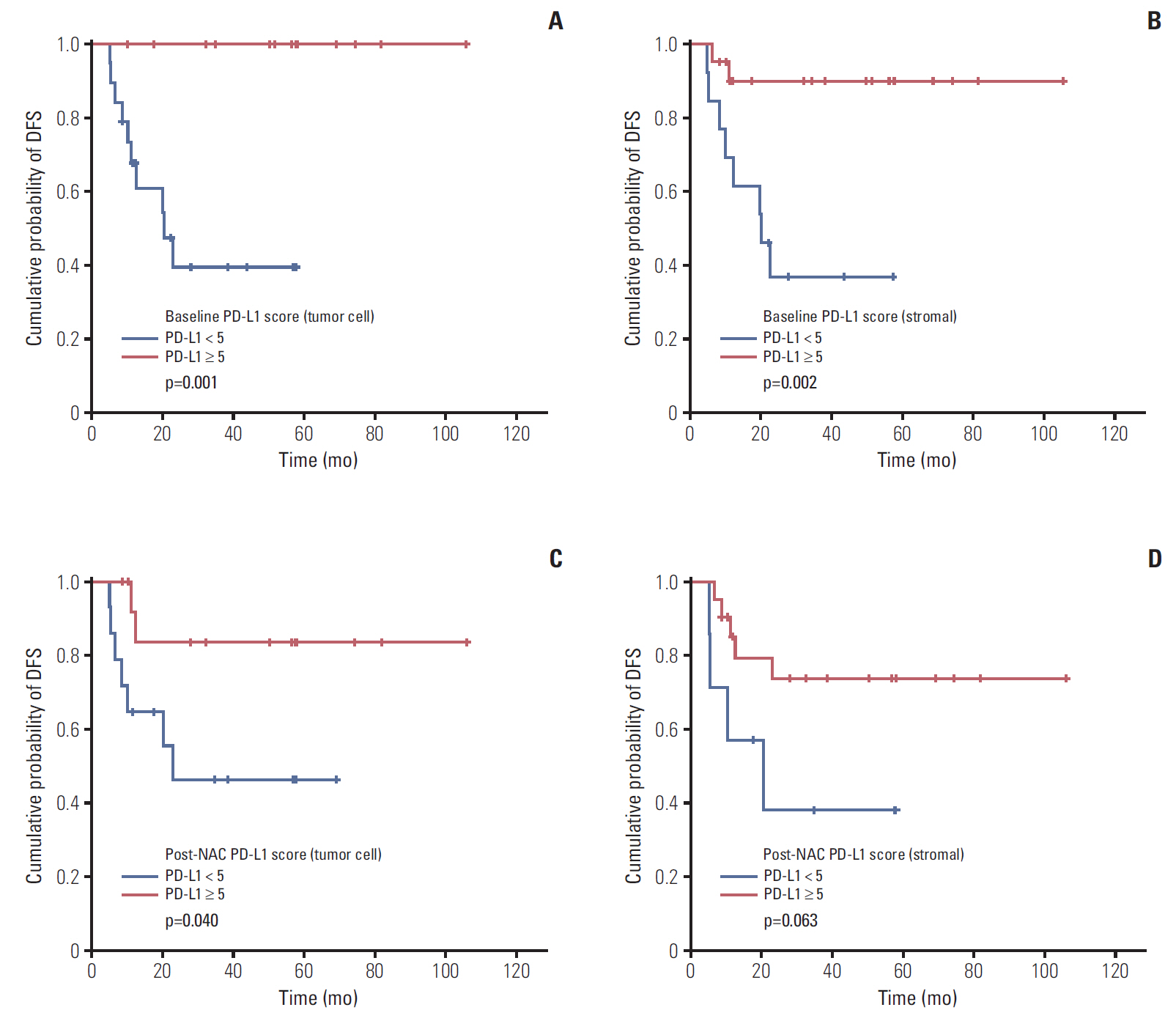
When analyzing baseline tumor tissue, positive tumor and stromal PD-L1 expression (PD-L1 H-score ≥ 5) was associated to superior DFS (tumor PD-L1 median DFS, 57.6 months vs. 12.5 months; p=0.001 and stromal PD-L1 median DFS, 50.17 months vs. 20.4 months; p=0.002) (Fig. 5A and B). After NAC, patients with positive tumor PD-L1 expression (H-score ≥ 5) showed longer DFS (median DFS, 53.27 months vs. 18.9 months; p=0.040) (Fig. 5C). In case of stromal PD-L1 expression after NAC, patients with H-score same or higher than 5 showed borderline significance for superior DFS compared to patient with H-score below 5 (median DFS, 38.37 months vs. 17.5 months; p=0.063) (Fig. 5D).
In our study, high baseline NLR was associated to poor survival outcomes in early and advanced TNBC, consistent with previous literatures [3,5]. NLR is thought to reflect inflammatory changes during cancer development [3] and has shown relationship with survival outcomes in various solid cancer [24]. There were reports about the role of NLR in TNBC in small population [3], and our study strengthened prior reports with relatively large number of patient population.
Until now, there were scarce reports about the role of changes of serum NLR connected to survival outcome in TNBC during chemotherapy. Considering NLR reflects inflammatory changes and survival outcomes in cancer progression, we hypothesized that changes of NLR during NAC might be associated to cancer response or survival during follow-up. In 50 patients who received NAC, radical changes of NLR (NLR change < –30% or > 100%) was associated with inferior DFS. The radical change of NLR was still associated to inferior DFS in multivariate analysis. Changes of NLR during NAC might have reflected cancer-mediated inflammatory changes during treatment [4] and connected to survival and prognosis of breast cancer. We initially assumed that increased NLR after NAC may be associated to poor survival or response, but subtle increase or decrease in serum NLR did not reflect survival outcome during analysis. Radical changes of serum NLR might be associated to definite changes in inflammatory response or changes of cancer environment during treatment, but further analysis should be warranted for confirmation of this hypothesis.
For the first time, we reported the relationship between serum NLR and tumor TIL during NAC. We presumed systemic inflammatory change may be associated to tumor inflammatory change during NAC, and for confirmation of this hypothesis, we analyzed the relationship between changes of serum NLR with tumor TIL change during NAC. Serum NLR and tumor TIL was selected to represent systemic inflammatory changes and local tumor inflammatory changes, each. There were trends for negative correlation between changes of TIL and serum NLR during NAC, but there was no statistical significance. We assumed other systemic and local inflammatory markers such as cytokines, transcription factors, acute-phase proteins should be considered during interpretation [4], and these confounding factors may have influenced borderline association between serum NLR and tumor TIL.
Previous literature reports the relationship of high pretreatment TIL associated with favorable survival in breast cancer [18], and our result also showed trends for favorable DFS in patients with higher baseline TIL with borderline statistical significance (data not shown). In our study, we investigated the changes between TIL during NAC and analyzed the relationship with survival outcomes. Although the result was not statistically significant, we did find out that there were trends for increased survival in patients with increased or same TIL compared to baseline TIL during NAC. This result is in concordance with previous report [19]. In other retrospective analysis, decreasing TIL was detected after NAC and this change was associated to increased pCR rate, but there was no further analysis about accordance to survival outcome with change of TIL [25]. Previous study patient population was heterogeneous in hormonal status and HER2 status, with small patient number. In our study, although we recruited only TNBC patients, but the population was small in number. The change of TIL during NAC seems to influence the tumor response or survival in breast cancer including TNBC, but further study is needed with large number of patients with homogeneous characteristics, and sufficient follow-up duration is needed.
Subtyping of TIL with CD4+, CD8+, and FOXP3+ was done and their association with PD-L1 expression in tumor and stromal TIL was performed in this study. As reported previously, CD4+, CD8+, and FOXP3+ were mutually associated with each other in baseline and post-NAC tissues in our study [17,26]. PD-L1 expression in baseline tissue and post-NAC tissue was statistically associated to TIL, and this result is consistent with prior study [14]. When TIL was subtyped with CD4+, CD8+, or FOXP3+, subtyped TIL was partially associated to PD-L1 expression in baseline and post-NAC tissue. CD4+, CD8+, FOXP3+ TIL all showed trends for positive correlation of PD-L1 expression in tumor tissue. The relationship between TIL subtype and PD-L1 expression showed heterogeneous association among previous reported literatures, with conflicting results in breast cancer (BC) [17,27,28]. Persistent, or high level of PD-L1 expression may induced CD8+ T cell exhaustion, and also may be related to CD4+ or FOXP3+ T cells which maintain immune response and expression of PD-L1 expression [29]. Considering there are negative relationship between regulatory T cell, effector T cell and PD-L1 expression, negative correlation between TIL subtype, especially CD8+ TIL and PD-L1 might be estimated, and there are reports supporting this view [17,30]. However, there are other reports supporting CD8+ T cell may recruit CD4+ or FOXP3+ regulatory T cells with PD-L1 by intrinsic immune mechanism [31], and this hypothesis is in consistence to our result. When interpreting the relationship between TIL, TIL subtype and PD-L1 expression, various tumor induced environmental change and immune mechanism may influence the expression of T cell and PD-L1 expression, and this might have influenced the conflicting reports about the TIL subtype and PD-L1 expression in BC.
As one of an immune modulatory marker and representing tumor microenvironment [14], the change of PD-L1 expression during NAC was analyzed in the study. Patients who showed negative tumor PD-L1 or negative conversion of tumor PD-L1 expression during NAC showed poor outcome. Furthermore, low PD-L1 expression in tumor or stromal tissue (H-score < 5) in baseline tumor or post-NAC tissue also showed poor DFS. To date, the role of PD-L1 expression and its association to clinical outcome is controversial. Part or studies report increased PD-L1 expression is associated to poor prognosis or aggressive clinical behavior in BC or other cancers [15,17,21,30], and there are other studies reporting PD-L1 expression associated to favorable outcome in solid cancers, consistent with our analysis [14,20,32]. This discrepancy may arise from the heterogeneous composition of various solid tumors in the study, and various definition of PD-L1 status among each study may have influenced the conflicting results of previous reports as well. Furthermore, different molecular profile and oncogenic mechanism between BC subtypes or other types of cancer may have influenced the role of PD-L1, which might have relatively different role between various cancers and different breast cancer subtypes. The merit of our study lies on relatively homogenous patient population, consisted of locally advanced TNBC treated with similar systemic chemotherapy. Considering TNBC is more immunogenic compared to other subtypes, PD-L1 and its associated molecular pathway may have different role compared to other BC subtypes.
In present study, there are some limitations to mention. First, we included TNBC patients diagnosed and treated until Dec 2015. We included recently diagnosed TNBC patients with adequate archival tumor tissue, but the follow-up duration was relatively short compared to other studies. Long-term follow up of the results of our study is needed for future study. Second, as we mentioned, although the subtype was homogeneous, the patient population with adequate tumor tissue was relatively small, requiring careful interpretation of the analysis. However, this study has its clinical value for selecting only TNBC patients with paired tumor samples. Based on our analysis, we are planning to review the change of NLR, TIL, and PD-L1 expression in large number of scale, comprising multicenter in Korea. Sufficient follow-up duration with large number of patient population is warranted for future study.
In conclusion, this study showed high baseline NLR associated to poor survival outcome in locally advanced TNBC patients. Among patients receiving NAC, radical change of serum NLR during chemotherapy was associated to poor prognosis. Increased TIL in post-NAC tumor tissue showed trends for superior DFS during follow-up, but it did not reach statistical significance during analysis. Finally, negative conversion of PD-L1 expression or persistent negative expression of PD-L1 in tumor tissue during NAC or low level of PD-L1 expression in baseline or post-NAC tumor tissue was associated to poor prognosis. For more confirmative results, larger sample size of patients with sufficient follow-up duration is warranted.
Supplementary materials are available at Cancer Research and Treatment website (http://www.e-crt.org).

Conflict of interest relevant to this article was not reported.

Acknowledgements
The present research has been supported by Korea Breast Cancer Foundation and Basic Science Research Program through the National Research Foundation of Korea funded by the Ministry of Science, ICT and Future Planning (NRF-2017R1D1A1B03034165).
Fig. 1.
Representative staining of tumor infiltrating-lymphocytes (TILs) and programmed death ligand-1 (PD-L1) expression in tumor specimens. (A) CD4+ TILs (×400). (B) CD8+ TILs (×400). (C) Forkhead box P3+ TILs (×400). (D) PD-L1 (×400, H-score 200). (E) PD-L1 (×400, H-score 75).
crt-2018-270f1.jpg
Fig. 2.
(A) Overall survival (OS) of patients according to neutrophil-lymphocyte ratio (NLR) level. (B) Disease-free survival of patients according to NLR level. (C) Disease-free survival (DFS) of patients who received neoadjuvant chemotherapy according to changes of NLR.
crt-2018-270f2.jpg
Fig. 3.
(A) Correlation between neutrophil-lymphocyte ratio change and tumor infiltrating-lymphocyte (TIL) change during neoadjuvant chemotherapy (NAC). (B) Correlation between TIL and programmed death ligand-1 (PD-L1) changes in primary tumor tissue. (C) Correlation between TIL and PD-L1 changes in post-NAC tissue.
crt-2018-270f3.jpg
Fig. 4.
(A) Disease-free survival (DFS) according to changes of tumor infiltrating lymphocyte during neoadjuvant chemotherapy (NAC). (B) DFS according to change of tumor cell programmed death ligand-1 (PD-L1) expression change during NAC. (C) DFS according to change of stromal cell PD-L1 expression during NAC.
crt-2018-270f4.jpg
Fig. 5.
Disease-free survival (DFS) according to tumor cell programmed death ligand-1 expression (PD-L1) (A) and stromal cell PD-L1 expression (B) in baseline tumor tissue. DFS according to tumor cell PD-L1 expression (C) and stromal cell PD-L1 expression (D) in post-neoadjuvant chemotherapy (post-NAC) tumor tissues.
crt-2018-270f5.jpg
Table 1.
Baseline patient characteristics
Total NLR ≤ 3.16 NLR > 3.16 p-value
No. of patients 358 313 (87.4) 45 (12.6)
NLR 1.76 (0.12-10.06) 1.64 (0.12-3.16) 3.95 (3.2-10.06)
Age (yr)
 Median (range) 51 (14-86) 51 (14-86) 48 (32-83) 0.970
 < 40 71 (19.8) 62 (17.3) 9 (2.5)
 ≥ 40 287 (80.2) 251 (70.1) 36 (10.1)
Stage
 I 127 (35.5) 113 (31.6) 14 (3.9) 0.340
 II 164 (45.8) 145 (40.5) 19 (5.3)
 III 67 (18.7) 55 (15.3) 12 (3.4)
Histology
 Invasive ductal carcinoma 300 (83.8) 260 (72.6) 40 (11.2) 0.460
 Invasive lobular carcinoma 6 (1.7) 6 (1.7) 0
 Medullary carcinoma 17 (4.7) 14 (3.9) 3 (0.8)
 Metaplastic carcinoma 17 (4.7) 16 (4.4) 1 (0.3)
 Others 4 (1.1) 4 (1.1) 0
Histologic grade
 Well-moderately differentiated 103 (28.8) 86 (24.0) 17 (4.8) 0.408
 Poorly differentiated 252 (70.4) 224 (62.6) 28 (7.8)
 Unknown 3 (0.8) 3 (0.8) 0
Ki-67
 < 20% 48 (13.4) 43 (12.0) 5 (1.4) 0.677
 ≥ 20% 303 (84.6) 265 (74.0) 38 (10.6)
 Unknown 7 (2.0) 5 (1.4) 2 (0.6)
Adjuvant chemotherapy 310 (86.6) 271 (75.7) 39 (10.9) 0.540
 AC followed by docetaxel 64 (17.9) 59 (16.4) 5 (1.5)
 AC 171 (47.8) 149 (41.6) 22 (6.2)
 DA 14 (3.9) 8 (2.2) 6 (1.7)
 CMF 47 (13.1) 42 (11.7) 5 (1.4)
 TC 11 (3.1) 10 (2.8) 1 (0.3)
 TAC 2 (0.6) 2 (0.6) 0
 FEC 1 (0.3) 1 (0.3) 0
Adjuvant radiation
 Yes 267 (74.6) 234 (65.4) 33 (9.2) 0.900
 No 91 (25.4) 79 (22.1) 12 (3.3)
Cancer recurrence 47 (13.1) 37 (10.3) 10 (2.8) 0.047
Death 36 (10.1) 26 (7.3) 10 (2.8) 0.004

Values are presented as median (range) or number (%). NLR, neutrophil-to-lymphocyte ratio; AC, adriamycin-cyclophosphamide; DA, docetaxel-adriamycin; CMF, cyclophosphamide–methotrexate–5-fluorouracil; TC, docetaxel-cyclophosphamide; TAC, docetaxel-adriamycin-cyclophosphamide; FEC, 5-fluorouracil–epirubicin–cyclophosphamide.

Table 2.
Baseline characteristics of patients receiving neoadjuvant chemotherapy
Total NLR ≤ 3.16 NLR > 3.16 p-value
No. of patients 50 42 (84) 8 (16) 0.430
Age (yr)
 Median (range) 43.5 (25-66) 43.5 (36-51) 0.600
 < 40 15 (30) 13 (26) 2 (4)
 ≥ 40 35 (70) 29 (58) 6 (12)
Clinical stage
 II 9 (18) 9 (18) 0 0.150
 III 41 (82) 33 (66) 8 (16)
Pathologic stage
 0 3 (6) 3 (6) 0 0.260
 I 7 (14) 6 (12) 1 (2)
 II 16 (32) 12 (24) 4 (8)
 III 14 (28) 11 (22) 3 (6)
Downstaging
 No 6 (12) 4 (8) 2 (4) 0.500
 Yes 44 (88) 38 (76) 6 (12)
pCR 5 (10) 5 (10) 0 0.300
Neoadjuvant chemotherapy
 AC 13 (26) 11 (22) 2 (4) 0.870
 EC 5 (10) 5 (10) 0
 DA 32 (64) 26 (52) 6 (12)
Adjuvant radiation
 Yes 36 (72) 29 (58) 7 (14) 0.400
 No 14 (28) 13 (26) 1 (2)
Cancer recurrence 9 (18) 6 (12) 3 (6) 0.500
Death 8 (16) 6 (12) 2 (4) 0.450

Values are presented as number (%) unless otherwise indicated. NLR, neutrophil-to-lymphocyte ratio; pCR, pathologic compete response; AC, adriamycin-cyclophosphamide; EC, epirubicin-cyclophosphamide; DA, docetaxel-adriamycin.

Table 3.
Univariate and multivariate analysis of disease-free survival and overall survival
Characteristic Overall survival
Disease-free survival
Univariate analysis
Multivariate analysis
Univariate analysis
Multivariate analysis
HR (95% CI) p-value HR (95% CI) p-value HR (95% CI) p-value HR (95% CI) p-value
Age (yr)
 ≥ 40 vs. < 40 1.167 (0.6-2.67) 0.720 - - 0.83 (0.42-1.63) 0.580 - -
Stage
 I 1 (reference) < 0.001 1 (reference) 0.114 1 (reference) < 0.001 1 (reference) < 0.001
 II vs. I 2.13 (0.84-5.45) 0.110 3.15 (1.03-9.68) 0.045 1.41 (0.67-2.97) 0.361 1.33 (0.61-2.95) 0.470
 III vs. I 5.97 (2.23-15.6) < 0.001 3.12 (0.96-10.01) 0.058 4.07 (1.91-8.72) < 0.001 4.0 (1.81-8.85) 0.001
NLR
 > 3.16 vs. ≤ 3.16 2.97 (1.43-6.17) 0.003 3.15 (1.3-7.5) 0.009 2.11 (1.05-4.25) 0.036 1.73 (0.83-3.6) 0.140
Ki-67 (%)
 ≥ 20 vs. < 20 3.11 (0.74-13.1) 0.120 3.68 (0.87-15.7) 0.070 1.003 (0.44-2.26) 0.994 - -
History of adjuvant chemotherapy
 Yes vs. no 1.57 (0.48-5.14) 0.460 - - 2.18 (0.68-7.04) 0.190 - -
Cancer recurrence
 Yes vs. no 55.9 (19.7-159.2) < 0.001 58.2 (19.76-171.4) < 0.001 - - - -

HR, hazard ratio; CI, confidence interval; NLR, neutrophil-to-lymphocyte ratio.

Table 4.
Univariate and Multivariate analysis of disease-free survival in neoadjuvant patients
Characteristic Univariate analysis
Multivariate analysis
Hazard ratio (95% CI) p-value Hazard ratio (95% CI) p-value
Age (yr)
 < 40 vs. ≥ 40 1.07 (0.33-3.42) 0.900 - -
Clinical stage
 II vs. III 3.76 (0.49-28.85) 0.200 - -
Surgical stage 0.034 0.027
 0 vs. I 0.51 (0.046-5.65) 0.590 0.37 (0.33-4.24) 0.430
 0 vs. II 0.5 (0.052-4.83) 0.550 0.39 (0.4-3.84) 0.420
 0 vs. III 2.74 (0.34-22.08) 0.340 2.22 (0.27-18.0) 0.450
Downstaging
 No vs. yes 0.55 (0.19-1.59) 0.270 - -
pCR
 No vs. yes 0.62 (0.08-4.7) 0.640 - -
Baseline NLR
 ≤ 3.16 vs. > 3.16 1.8 (0.5-6.5) 0.360 - -
NLR change (%)
 –30% < NLR change < 100% vs. NLR < –30% or > 100% 3.81 (1.19-12.15) 0.024 4.1 (1.27-13.16) 0.018
Ki-67 (%)
 < 20 vs. ≥ 20 1.29 (0.36-4.64) 0.690 - -

CI, confidence interval; pCR, pathologic complete response; NLR, neutrophil-to-lymphocyte ratio.

Table 5.
Distribution of TILs and PD-L1 in tumor tissues
Baseline tumor tissue Tumor tissue after neoadjuvant chemotherapy
No. of patients 34 30
Distribution of sTIL 10 (3-35) 11.5 (5-60)
CD4 74.6 (9.8-226) 67.8 (11.6-250.6)
CD8 61.2 (6-166.8) 85 (19.6-189)
FOXP3 12.4 (1.8-54.6) 10.2 (1-41)
CD8/FOXP3 ratio 4.21 (1.2-44.9) 7.66 (2.96-26.4)
PD-L1 (tumor cell)
 Median (range) 1.5 (0-200) 2 (0-270)
 ≥ 1 20 (58.8) 17 (56.7)
 ≥ 5 15 (44.1) 14 (46.7)
PD-L1 (stromal cell)
 Median (range) 8.5 (0-90) 17.5 (0-125)
 ≥ 1 30 (88.2) 24 (80.0)
 ≥ 5 21 (61.8) 21 (70.0)
TIL change during NAC
 Same or increased 21 (70.0)
 Decreased 9 (30.0)
TIL fold change during NAC
 Median (range) 1.25 (0.5-8.33)
PD-L1 change during NAC
 Negative or negative conversion in tumor 12 (35.3)
 Negative or negative conversion in stroma 4 (13.3)

Value are presented as median (range) or number (%). TIL, tumor-infiltrating lymphocyte; PD-L1, programmed death-ligand 1; sTIL, stromal TIL; FOXP3, forkhead box P3; NAC, neoadjuvant chemotherapy.

  • 1. Malorni L, Shetty PB, De Angelis C, Hilsenbeck S, Rimawi MF, Elledge R, et al. Clinical and biologic features of triple-negative breast cancers in a large cohort of patients with long-term follow-up. Breast Cancer Res Treat. 2012;136:795–804. ArticlePubMedPMC
  • 2. Lehmann BD, Bauer JA, Chen X, Sanders ME, Chakravarthy AB, Shyr Y, et al. Identification of human triple-negative breast cancer subtypes and preclinical models for selection of targeted therapies. J Clin Invest. 2011;121:2750–67. ArticlePubMedPMC
  • 3. Pistelli M, De Lisa M, Ballatore Z, Caramanti M, Pagliacci A, Battelli N, et al. Pre-treatment neutrophil to lymphocyte ratio may be a useful tool in predicting survival in early triple negative breast cancer patients. BMC Cancer. 2015;15:195.ArticlePubMedPMCPDF
  • 4. Diakos CI, Charles KA, McMillan DC, Clarke SJ. Cancer-related inflammation and treatment effectiveness. Lancet Oncol. 2014;15:e493–503. ArticlePubMed
  • 5. Orditura M, Galizia G, Diana A, Saccone C, Cobellis L, Ventriglia J, et al. Neutrophil to lymphocyte ratio (NLR) for prediction of distant metastasis-free survival (DMFS) in early breast cancer: a propensity score-matched analysis. ESMO Open. 2016;1:e000038ArticlePubMedPMC
  • 6. Azab B, Bhatt VR, Phookan J, Murukutla S, Kohn N, Terjanian T, et al. Usefulness of the neutrophil-to-lymphocyte ratio in predicting short- and long-term mortality in breast cancer patients. Ann Surg Oncol. 2012;19:217–24. ArticlePubMed
  • 7. Chen Y, Chen K, Xiao X, Nie Y, Qu S, Gong C, et al. Pretreatment neutrophil-to-lymphocyte ratio is correlated with response to neoadjuvant chemotherapy as an independent prognostic indicator in breast cancer patients: a retrospective study. BMC Cancer. 2016;16:320.ArticlePubMedPMCPDF
  • 8. Loi S, Sirtaine N, Piette F, Salgado R, Viale G, Van Eenoo F, et al. Prognostic and predictive value of tumor-infiltrating lymphocytes in a phase III randomized adjuvant breast cancer trial in node-positive breast cancer comparing the addition of docetaxel to doxorubicin with doxorubicin-based chemotherapy: BIG 02-98. J Clin Oncol. 2013;31:860–7. ArticlePubMed
  • 9. Zhang L, Conejo-Garcia JR, Katsaros D, Gimotty PA, Massobrio M, Regnani G, et al. Intratumoral T cells, recurrence, and survival in epithelial ovarian cancer. N Engl J Med. 2003;348:203–13. ArticlePubMed
  • 10. Galon J, Costes A, Sanchez-Cabo F, Kirilovsky A, Mlecnik B, Lagorce-Pages C, et al. Type, density, and location of immune cells within human colorectal tumors predict clinical outcome. Science. 2006;313:1960–4. ArticlePubMed
  • 11. Miyashita M, Sasano H, Tamaki K, Hirakawa H, Takahashi Y, Nakagawa S, et al. Prognostic significance of tumor-infiltrating CD8+ and FOXP3+ lymphocytes in residual tumors and alterations in these parameters after neoadjuvant chemotherapy in triple-negative breast cancer: a retrospective multicenter study. Breast Cancer Res. 2015;17:124.ArticlePubMedPMC
  • 12. Chen DS, Irving BA, Hodi FS. Molecular pathways: next-generation immunotherapy: inhibiting programmed death-ligand 1 and programmed death-1. Clin Cancer Res. 2012;18:6580–7. ArticlePubMed
  • 13. Patel SP, Kurzrock R. PD-L1 expression as a predictive biomarker in cancer immunotherapy. Mol Cancer Ther. 2015;14:847–56. ArticlePubMed
  • 14. Wimberly H, Brown JR, Schalper K, Haack H, Silver MR, Nixon C, et al. PD-L1 expression correlates with tumor-infiltrating lymphocytes and response to neoadjuvant chemotherapy in breast cancer. Cancer Immunol Res. 2015;3:326–32. ArticlePubMed
  • 15. Muenst S, Schaerli AR, Gao F, Daster S, Trella E, Droeser RA, et al. Expression of programmed death ligand 1 (PD-L1) is associated with poor prognosis in human breast cancer. Breast Cancer Res Treat. 2014;146:15–24. ArticlePubMedPMC
  • 16. Sabatier R, Finetti P, Mamessier E, Adelaide J, Chaffanet M, Ali HR, et al. Prognostic and predictive value of PD-L1 expression in breast cancer. Oncotarget. 2015;6:5449–64. ArticlePubMed
  • 17. Park IH, Kong SY, Ro JY, Kwon Y, Kang JH, Mo HJ, et al. Prognostic implications of tumor-infiltrating lymphocytes in association with programmed death ligand 1 expression in early-stage breast cancer. Clin Breast Cancer. 2016;16:51–8. ArticlePubMed
  • 18. Pruneri G, Vingiani A, Bagnardi V, Rotmensz N, De Rose A, Palazzo A, et al. Clinical validity of tumor-infiltrating lymphocytes analysis in patients with triple-negative breast cancer. Ann Oncol. 2016;27:249–56. ArticlePubMedPDF
  • 19. Pelekanou V, Carvajal-Hausdorf DE, Altan M, Wasserman B, Carvajal-Hausdorf C, Wimberly H, et al. Effect of neoadjuvant chemotherapy on tumor-infiltrating lymphocytes and PD-L1 expression in breast cancer and its clinical significance. Breast Cancer Res. 2017;19:91.ArticlePubMedPMCPDF
  • 20. Li X, Wetherilt CS, Krishnamurti U, Yang J, Ma Y, Styblo TM, et al. Stromal PD-L1 expression is associated with better disease-free survival in triple-negative breast cancer. Am J Clin Pathol. 2016;146:496–502. ArticlePubMedPDF
  • 21. Muenst S, Soysal SD, Gao F, Obermann EC, Oertli D, Gillanders WE. The presence of programmed death 1 (PD-1)-positive tumor-infiltrating lymphocytes is associated with poor prognosis in human breast cancer. Breast Cancer Res Treat. 2013;139:667–76. ArticlePubMed
  • 22. Hammond ME, Hayes DF, Dowsett M, Allred DC, Hagerty KL, Badve S, et al. American Society of Clinical Oncology/College of American Pathologists guideline recommendations for immunohistochemical testing of estrogen and progesterone receptors in breast cancer (unabridged version). Arch Pathol Lab Med. 2010;134:e48–72. ArticlePubMedPDF
  • 23. Wolff AC, Hammond ME, Hicks DG, Dowsett M, McShane LM, Allison KH, et al. Recommendations for human epidermal growth factor receptor 2 testing in breast cancer: American Society of Clinical Oncology/College of American Pathologists clinical practice guideline update. Arch Pathol Lab Med. 2014;138:241–56. ArticlePubMed
  • 24. Guthrie GJ, Charles KA, Roxburgh CS, Horgan PG, McMillan DC, Clarke SJ. The systemic inflammation-based neutrophil-lymphocyte ratio: experience in patients with cancer. Crit Rev Oncol Hematol. 2013;88:218–30. ArticlePubMed
  • 25. Pelekanou V, Barlow WE, von Wahlde MK, Wasserman B, Lo YC, Hayes DF, et al. Effects of neoadjuvant chemotherapy (NAC) on tumor infiltrating lymphocytes (TIL) and PD-L1 expression in the SWOG S0800 clinical trial. J Clin Oncol. 2017;35(15 Suppl):519.Article
  • 26. Seo AN, Lee HJ, Kim EJ, Kim HJ, Jang MH, Lee HE, et al. Tumour-infiltrating CD8+ lymphocytes as an independent predictive factor for pathological complete response to primary systemic therapy in breast cancer. Br J Cancer. 2013;109:2705–13. ArticlePubMedPMCPDF
  • 27. Cimino-Mathews A, Thompson E, Taube JM, Ye X, Lu Y, Meeker A, et al. PD-L1 (B7-H1) expression and the immune tumor microenvironment in primary and metastatic breast carcinomas. Hum Pathol. 2016;47:52–63. ArticlePubMed
  • 28. Ghebeh H, Barhoush E, Tulbah A, Elkum N, Al-Tweigeri T, Dermime S. FOXP3+ Tregs and B7-H1+/PD-1+ T lymphocytes co-infiltrate the tumor tissues of high-risk breast cancer patients: Implication for immunotherapy. BMC Cancer. 2008;8:57.ArticlePubMedPMCPDF
  • 29. Ohaegbulam KC, Assal A, Lazar-Molnar E, Yao Y, Zang X. Human cancer immunotherapy with antibodies to the PD-1 and PD-L1 pathway. Trends Mol Med. 2015;21:24–33. ArticlePubMed
  • 30. Hamanishi J, Mandai M, Iwasaki M, Okazaki T, Tanaka Y, Yamaguchi K, et al. Programmed cell death 1 ligand 1 and tumor-infiltrating CD8+ T lymphocytes are prognostic factors of human ovarian cancer. Proc Natl Acad Sci U S A. 2007;104:3360–5. ArticlePubMedPMC
  • 31. Spranger S, Spaapen RM, Zha Y, Williams J, Meng Y, Ha TT, et al. Up-regulation of PD-L1, IDO, and T(regs) in the melanoma tumor microenvironment is driven by CD8(+) T cells. Sci Transl Med. 2013;5:200ra116.ArticlePubMedPMC
  • 32. Badoual C, Hans S, Merillon N, Van Ryswick C, Ravel P, Benhamouda N, et al. PD-1-expressing tumor-infiltrating T cells are a favorable prognostic biomarker in HPV-associated head and neck cancer. Cancer Res. 2013;73:128–38. ArticlePubMed

Figure & Data

REFERENCES

    Citations

    Citations to this article as recorded by  
    • Neutrophil–lymphocyte ratio reflects tumour‐infiltrating lymphocytes and tumour‐associated macrophages and independently predicts poor outcome in breast cancers with neoadjuvant chemotherapy
      Joshua J X Li, Shelly Y B Ni, Julia Y S Tsang, Wai Yin Chan, Ray K W Hung, Joshua W H Lui, Sally W Y Ng, Leong Kwong Shum, Ying Fei Tang, Gary M Tse
      Histopathology.2024; 84(5): 810.     CrossRef
    • Circulating blood biomarkers correlated with the prognosis of advanced triple negative breast cancer
      Xingyu Li, Yanyan Zhang, Cheng Zhu, Wentao Xu, Xiaolei Hu, Domingo Antonio Sánchez Martínez, José Luis Alonso Romero, Ming Yan, Ying Dai, Hua Wang
      BMC Women's Health.2024;[Epub]     CrossRef
    • Recent advancements in nanoconstructs for the theranostics applications for triple negative breast cancer
      Ashutosh Gupta, Kumar Nishchaya, Moumita Saha, Gaurisha Alias Resha Ramnath Naik, Sarika Yadav, Shreya Srivastava, Amrita Arup Roy, Sudheer Moorkoth, Srinivas Mutalik, Namdev Dhas
      Journal of Drug Delivery Science and Technology.2024; 93: 105401.     CrossRef
    • Predictive value of stromal tumor-infiltrating lymphocytes in patients with breast cancer treated with neoadjuvant chemotherapy: A meta-analysis
      Guangfa Xia, Ziran Zhang, Qin Jiang, Huan Wang, Jie Wang
      Medicine.2024; 103(6): e36810.     CrossRef
    • The prognostic value of preoperative neoindices consisting of lymphocytes, neutrophils and albumin (LANR) in operable breast cancer: a retrospective study
      Yuan Wang, Jiaru Zhuang, Shan Wang, Yibo Wu, Ling Chen
      PeerJ.2024; 12: e17382.     CrossRef
    • A review concerning the breast cancer-related tumour microenvironment
      Oscar Hernán Rodríguez-Bejarano, Carlos Parra-López, Manuel Alfonso Patarroyo
      Critical Reviews in Oncology/Hematology.2024; 199: 104389.     CrossRef
    • Alteration of PD-L1 (SP142) status after neoadjuvant chemotherapy and its clinical significance in triple-negative breast cancer
      Ji Won Woo, Eun Kyung Han, Koung Jin Suh, Se Hyun Kim, Jee Hyun Kim, So Yeon Park
      Breast Cancer Research and Treatment.2024; 207(2): 301.     CrossRef
    • Predictive value of pretreatment circulating inflammatory response markers in the neoadjuvant treatment of breast cancer: meta-analysis
      Gavin P Dowling, Gordon R Daly, Aisling Hegarty, Sandra Hembrecht, Aisling Bracken, Sinead Toomey, Bryan T Hennessy, Arnold D K Hill
      British Journal of Surgery.2024;[Epub]     CrossRef
    • Pathology of neoadjuvant therapy and immunotherapy testing for breast cancer
      Elena Provenzano, Abeer M Shaaban
      Histopathology.2023; 82(1): 170.     CrossRef
    • Recent Advances in Targeted Nanocarriers for the Management of Triple Negative Breast Cancer
      Rajesh Pradhan, Anuradha Dey, Rajeev Taliyan, Anu Puri, Sanskruti Kharavtekar, Sunil Kumar Dubey
      Pharmaceutics.2023; 15(1): 246.     CrossRef
    • The prognostic value of tumour-infiltrating lymphocytes, programmed cell death protein-1 and programmed cell death ligand-1 in Stage I–III triple-negative breast cancer
      Guang-Yi Sun, Jing Zhang, Bing-Zhi Wang, Hao Jing, Hui Fang, Yu Tang, Yong-Wen Song, Jing Jin, Yue-Ping Liu, Yuan Tang, Shu-Nan Qi, Bo Chen, Ning-Ning Lu, Ning Li, Ye-Xiong Li, Jian-Ming Ying, Shu-Lian Wang
      British Journal of Cancer.2023; 128(11): 2044.     CrossRef
    • Effect of neoadjuvant chemotherapy on tumor immune infiltration in breast cancer patients: Systematic review and meta-analysis
      Manuela Llano-León, Laura Camila Martínez-Enriquez, Oscar Mauricio Rodríguez-Bohórquez, Esteban Alejandro Velandia-Vargas, Nicolás Lalinde-Ruíz, María Alejandra Villota-Álava, Ivon Johanna Rodríguez-Rodríguez, María del Pilar Montilla-Velásquez, Carlos Al
      PLOS ONE.2023; 18(4): e0277714.     CrossRef
    • Association of neutrophil-to-lymphocyte ratio with clinical, pathological, radiological, laboratory features and disease outcomes of invasive breast cancer patients: A retrospective observational cohort study
      Sarosh Khan Jadoon, Rufina Soomro, Muhammad Nadeem Ahsan, Raja Muhammad Ijaz Khan, Sadia Iqbal, Farah Yasmin, Hala Najeeb, Nida Saleem, Namiya Cho, Resham, Taha Gul Shaikh, Syeda Fatima Saba Hasan, Muhammad Zain Khalid, Sarosh Alvi, Ahsan Mujtaba Rizvi,
      Medicine.2023; 102(20): e33811.     CrossRef
    • A new prognostic model including immune biomarkers, genomic proliferation tumor markers (AURKA and MYBL2) and clinical-pathological features optimizes prognosis in neoadjuvant breast cancer patients
      Esmeralda García-Torralba, Esther Navarro Manzano, Gines Luengo-Gil, Pilar De la Morena Barrio, Asunción Chaves Benito, Miguel Pérez-Ramos, Beatriz Álvarez-Abril, Alejandra Ivars Rubio, Elisa García-Garre, Francisco Ayala de la Peña, Elena García-Martínez
      Frontiers in Oncology.2023;[Epub]     CrossRef
    • Tumor infiltrating lymphocytes and neutrophil-to-lymphocyte ratio in relation to pathological complete remission to neoadjuvant therapy and prognosis in triple negative breast cancer
      Meng Zhao, Hui Xing, Jiankun He, Xinran Wang, Yueping Liu
      Pathology - Research and Practice.2023; 248: 154687.     CrossRef
    • Minimal residual disease in advanced or metastatic solid cancers: The G0-G1 state and immunotherapy are key to unwinding cancer complexity
      Andrea Nicolini, Giuseppe Rossi, Paola Ferrari, Angelo Carpi
      Seminars in Cancer Biology.2022; 79: 68.     CrossRef
    • The advanced lung cancer inflammation index is a novel independent prognosticator in colorectal cancer patients after curative resection
      Taichi Horino, Ryuma Tokunaga, Yuji Miyamoto, Yukiharu Hiyoshi, Takahiko Akiyama, Nobuya Daitoku, Yuki Sakamoto, Naoya Yoshida, Hideo Baba
      Annals of Gastroenterological Surgery.2022; 6(1): 83.     CrossRef
    • The Characteristics of Tumor Microenvironment in Triple Negative Breast Cancer
      Yiqi Fan, Shuai He
      Cancer Management and Research.2022; Volume 14: 1.     CrossRef
    • Predictive and Prognostic Role of Peripheral Blood T-Cell Subsets in Triple-Negative Breast Cancer
      Meng Li, Junnan Xu, Cui Jiang, Jingyan Zhang, Tao Sun
      Frontiers in Oncology.2022;[Epub]     CrossRef
    • PD-L1 expression in breast invasive ductal carcinoma with incomplete pathological response to neoadjuvant chemotherapy
      Ahmad Alhesa, Heyam Awad, Sarah Bloukh, Mahmoud Al-Balas, Mohammed El-Sadoni, Duaa Qattan, Bilal Azab, Tareq Saleh
      International Journal of Immunopathology and Pharmacology.2022;[Epub]     CrossRef
    • Circulating proteins as predictive and prognostic biomarkers in breast cancer
      Hugo Veyssière, Yannick Bidet, Frederique Penault-Llorca, Nina Radosevic-Robin, Xavier Durando
      Clinical Proteomics.2022;[Epub]     CrossRef
    • Targeting the antigen processing and presentation pathway to overcome resistance to immune checkpoint therapy
      Silvia D’Amico, Patrizia Tempora, Ombretta Melaiu, Valeria Lucarini, Loredana Cifaldi, Franco Locatelli, Doriana Fruci
      Frontiers in Immunology.2022;[Epub]     CrossRef
    • Programmed death-ligand 1 (PD-L1) expression predicts response to neoadjuvant chemotherapy in triple-negative breast cancer: a systematic review and meta-analysis
      Hamdy A. Azim, Kyrillus S. Shohdy, Hagar Elghazawy, Monica M. Salib, Doaa Almeldin, Loay Kassem
      Biomarkers.2022; 27(8): 764.     CrossRef
    • Determining Factors in the Therapeutic Success of Checkpoint Immunotherapies against PD-L1 in Breast Cancer: A Focus on Epithelial-Mesenchymal Transition Activation
      Mariana Segovia-Mendoza, Susana Romero-Garcia, Cristina Lemini, Heriberto Prado-Garcia, francesca bianchi
      Journal of Immunology Research.2021; 2021: 1.     CrossRef
    • Kaiso (ZBTB33) subcellular partitioning functionally links LC3A/B, the tumor microenvironment, and breast cancer survival
      Sandeep K. Singhal, Jung S. Byun, Samson Park, Tingfen Yan, Ryan Yancey, Ambar Caban, Sara Gil Hernandez, Stephen M. Hewitt, Heike Boisvert, Stephanie Hennek, Mark Bobrow, Md Shakir Uddin Ahmed, Jason White, Clayton Yates, Andrew Aukerman, Rami Vanguri, R
      Communications Biology.2021;[Epub]     CrossRef
    • Relationship Between the Neutrophil to Lymphocyte Ratio, Stromal Tumor-infiltrating Lymphocytes, and the Prognosis and Response to Neoadjuvant Chemotherapy in Triple-negative Breast Cancer
      Jian Pang, Haiyan Zhou, Xue Dong, Shouman Wang, Zhi Xiao
      Clinical Breast Cancer.2021; 21(6): e681.     CrossRef
    • Immune cell composition and functional marker dynamics from multiplexed immunohistochemistry to predict response to neoadjuvant chemotherapy in the WSG-ADAPT-TN trial
      Monika Graeser, Friedrich Feuerhake, Oleg Gluz, Valery Volk, Michael Hauptmann, Katarzyna Jozwiak, Matthias Christgen, Sherko Kuemmel, Eva-Maria Grischke, Helmut Forstbauer, Michael Braun, Mathias Warm, John Hackmann, Christoph Uleer, Bahriye Aktas, Claud
      Journal for ImmunoTherapy of Cancer.2021; 9(5): e002198.     CrossRef
    • Clinicopathological and prognostic significance of programmed cell death ligand 1 expression in patients diagnosed with breast cancer: meta-analysis
      M G Davey, É J Ryan, M S Davey, A J Lowery, N Miller, M J Kerin
      British Journal of Surgery.2021; 108(6): 622.     CrossRef
    • INSTIGO Trial: Evaluation of a Plasma Protein Profile as a Predictive Biomarker for Metastatic Relapse of Triple Negative Breast Cancer
      Hugo Veyssière, Sejdi Lusho, Ioana Molnar, Myriam Kossai, Maureen Bernadach, Catherine Abrial, Yannick Bidet, Nina Radosevic-Robin, Xavier Durando
      Frontiers in Oncology.2021;[Epub]     CrossRef
    • Inhibitors of PD-1/PD-L1 and ERK1/2 impede the proliferation of receptor positive and triple-negative breast cancer cell lines
      Karen Bräutigam, Elodie Kabore-Wolff, Ahmad Fawzi Hussain, Stephan Polack, Achim Rody, Lars Hanker, Frank Köster
      Journal of Cancer Research and Clinical Oncology.2021; 147(10): 2923.     CrossRef
    • Prognostic Relevance of Neutrophil to Lymphocyte Ratio (NLR) in Luminal Breast Cancer: A Retrospective Analysis in the Neoadjuvant Setting
      Antonino Grassadonia, Vincenzo Graziano, Laura Iezzi, Patrizia Vici, Maddalena Barba, Laura Pizzuti, Giuseppe Cicero, Eriseld Krasniqi, Marco Mazzotta, Daniele Marinelli, Antonella Amodio, Clara Natoli, Nicola Tinari
      Cells.2021; 10(7): 1685.     CrossRef
    • Time-Sequencing of the Neutrophil-to-Lymphocyte Ratio to Predict Prognosis of Triple-Negative Breast Cancer
      Joo-Heung Kim, Nak-Hoon Son, Jun-Sang Lee, Ji-Eun Mun, Jee-Ye Kim, Hyung-Seok Park, Seho Park, Seung-Il Kim, Byeong-Woo Park
      Cancers.2021; 13(14): 3472.     CrossRef
    • Platelet-to-Lymphocyte Ratio Is Associated With Favorable Response to Neoadjuvant Chemotherapy in Triple Negative Breast Cancer: A Study on 120 Patients
      Sejdi Lusho, Xavier Durando, Marie-Ange Mouret-Reynier, Myriam Kossai, Nathalie Lacrampe, Ioana Molnar, Frederique Penault-Llorca, Nina Radosevic-Robin, Catherine Abrial
      Frontiers in Oncology.2021;[Epub]     CrossRef
    • Role of neutrophil-to-lymphocyte ratio as a prognostic biomarker in patients with breast cancer receiving neoadjuvant chemotherapy: a meta-analysis
      Qiong Zhou, Jie Dong, Qingqing Sun, Nannan Lu, Yueyin Pan, Xinghua Han
      BMJ Open.2021; 11(9): e047957.     CrossRef
    • Neutrophil to Lymphocyte Ratio after Treatment Completion as a Potential Predictor of Survival in Patients with Triple-Negative Breast Cancer
      Kwang-Min Kim, Hyang Suk Choi, Hany Noh, In-Jeong Cho, Seung Taek Lim, Jong-In Lee, Airi Han
      Journal of Breast Cancer.2021; 24(5): 443.     CrossRef
    • Tumor Microenvironment Characterization in Breast Cancer Identifies Prognostic and Neoadjuvant Chemotherapy Relevant Signatures
      Fei Ji, Jiao-Mei Yuan, Hong-Fei Gao, Ai-Qi Xu, Zheng Yang, Ci-Qiu Yang, Liu-Lu Zhang, Mei Yang, Jie-Qing Li, Teng Zhu, Min-Yi Cheng, Si-Yan Wu, Kun Wang
      Frontiers in Molecular Biosciences.2021;[Epub]     CrossRef
    • Neutrophil to Lymphocyte Ratio as Prognostic and Predictive Factor in Breast Cancer Patients: A Systematic Review
      Iléana Corbeau, William Jacot, Séverine Guiu
      Cancers.2020; 12(4): 958.     CrossRef
    • A Rosetta Stone for Breast Cancer: Prognostic Value and Dynamic Regulation of Neutrophil in Tumor Microenvironment
      Wei Zhang, Yimin Shen, Huanhuan Huang, Sheng Pan, Jingxin Jiang, Wuzhen Chen, Ting Zhang, Chao Zhang, Chao Ni
      Frontiers in Immunology.2020;[Epub]     CrossRef
    • Perspectives on Triple-Negative Breast Cancer: Current Treatment Strategies, Unmet Needs, and Potential Targets for Future Therapies
      Gagan K. Gupta, Amber L. Collier, Dasom Lee, Richard A. Hoefer, Vasilena Zheleva, Lauren L. Siewertsz van Reesema, Angela M. Tang-Tan, Mary L. Guye, David Z. Chang, Janet S. Winston, Billur Samli, Rick J. Jansen, Emanuel F. Petricoin, Matthew P. Goetz, Ha
      Cancers.2020; 12(9): 2392.     CrossRef
    • Prognostic Role and Clinical Significance of Tumor-Infiltrating Lymphocyte (TIL) and Programmed Death Ligand 1 (PD-L1) Expression in Triple-Negative Breast Cancer (TNBC): A Systematic Review and Meta-Analysis Study
      Parisa Lotfinejad, Mohammad Asghari Jafarabadi, Mahdi Abdoli Shadbad, Tohid Kazemi, Fariba Pashazadeh, Siamak Sandoghchian Shotorbani, Farhad Jadidi Niaragh, Amir Baghbanzadeh, Nafiseh Vahed, Nicola Silvestris, Behzad Baradaran
      Diagnostics.2020; 10(9): 704.     CrossRef
    • Prediction of Late Recurrence and Distant Metastasis in Early-stage Breast Cancer: Overview of Current and Emerging Biomarkers
      A. Gouri, B. Benarba, A. Dekaken, H. Aoures, S. Benharkat
      Current Drug Targets.2020; 21(10): 1008.     CrossRef
    • Prognostic value of neutrophil‐to‐lymphocyte ratio and platelet‐to‐lymphocyte ratio for breast cancer patients: An updated meta‐analysis of 17079 individuals
      Wanying Guo, Xin Lu, Qipeng Liu, Ting Zhang, Peng Li, Weiqiang Qiao, Miao Deng
      Cancer Medicine.2019; 8(9): 4135.     CrossRef
    • Prognostic and clinicopathological value of PD-L1 expression in primary breast cancer: a meta-analysis
      Wenfa Huang, Ran Ran, Bin Shao, Huiping Li
      Breast Cancer Research and Treatment.2019; 178(1): 17.     CrossRef
    • Comparison of Preoperative Neutrophil-Lymphocyte and Platelet-Lymphocyte Ratios in Bladder Cancer Patients Undergoing Radical Cystectomy
      Ruiliang Wang, Yang Yan, Shenghua Liu, Xudong Yao
      BioMed Research International.2019; 2019: 1.     CrossRef
    • Immunotherapy in HER2-positive breast cancer: state of the art and future perspectives
      E. Krasniqi, G. Barchiesi, L. Pizzuti, M. Mazzotta, A. Venuti, M. Maugeri-Saccà, G. Sanguineti, G. Massimiani, D. Sergi, S. Carpano, P. Marchetti, S. Tomao, T. Gamucci, R. De Maria, F. Tomao, C. Natoli, N. Tinari, G. Ciliberto, M. Barba, P. Vici
      Journal of Hematology & Oncology.2019;[Epub]     CrossRef
    • Prognostic Implications of PD-L1 Expression in Breast Cancer: Systematic Review and Meta-analysis of Immunohistochemistry and Pooled Analysis of Transcriptomic Data
      Alexios Matikas, Ioannis Zerdes, John Lövrot, François Richard, Christos Sotiriou, Jonas Bergh, Antonios Valachis, Theodoros Foukakis
      Clinical Cancer Research.2019; 25(18): 5717.     CrossRef

    • PubReader PubReader
    • ePub LinkePub Link
    • Cite
      CITE
      export Copy Download
      Close
      Download Citation
      Download a citation file in RIS format that can be imported by all major citation management software, including EndNote, ProCite, RefWorks, and Reference Manager.

      Format:
      • RIS — For EndNote, ProCite, RefWorks, and most other reference management software
      • BibTeX — For JabRef, BibDesk, and other BibTeX-specific software
      Include:
      • Citation for the content below
      Prognostic Role and Clinical Association of Tumor-Infiltrating Lymphocyte, Programmed Death Ligand-1 Expression with Neutrophil-Lymphocyte Ratio in Locally Advanced Triple-Negative Breast Cancer
      Cancer Res Treat. 2019;51(2):649-663.   Published online July 31, 2018
      Close
    • XML DownloadXML Download
    Prognostic Role and Clinical Association of Tumor-Infiltrating Lymphocyte, Programmed Death Ligand-1 Expression with Neutrophil-Lymphocyte Ratio in Locally Advanced Triple-Negative Breast Cancer
    Image Image Image Image Image
    Fig. 1. Representative staining of tumor infiltrating-lymphocytes (TILs) and programmed death ligand-1 (PD-L1) expression in tumor specimens. (A) CD4+ TILs (×400). (B) CD8+ TILs (×400). (C) Forkhead box P3+ TILs (×400). (D) PD-L1 (×400, H-score 200). (E) PD-L1 (×400, H-score 75).
    Fig. 2. (A) Overall survival (OS) of patients according to neutrophil-lymphocyte ratio (NLR) level. (B) Disease-free survival of patients according to NLR level. (C) Disease-free survival (DFS) of patients who received neoadjuvant chemotherapy according to changes of NLR.
    Fig. 3. (A) Correlation between neutrophil-lymphocyte ratio change and tumor infiltrating-lymphocyte (TIL) change during neoadjuvant chemotherapy (NAC). (B) Correlation between TIL and programmed death ligand-1 (PD-L1) changes in primary tumor tissue. (C) Correlation between TIL and PD-L1 changes in post-NAC tissue.
    Fig. 4. (A) Disease-free survival (DFS) according to changes of tumor infiltrating lymphocyte during neoadjuvant chemotherapy (NAC). (B) DFS according to change of tumor cell programmed death ligand-1 (PD-L1) expression change during NAC. (C) DFS according to change of stromal cell PD-L1 expression during NAC.
    Fig. 5. Disease-free survival (DFS) according to tumor cell programmed death ligand-1 expression (PD-L1) (A) and stromal cell PD-L1 expression (B) in baseline tumor tissue. DFS according to tumor cell PD-L1 expression (C) and stromal cell PD-L1 expression (D) in post-neoadjuvant chemotherapy (post-NAC) tumor tissues.
    Prognostic Role and Clinical Association of Tumor-Infiltrating Lymphocyte, Programmed Death Ligand-1 Expression with Neutrophil-Lymphocyte Ratio in Locally Advanced Triple-Negative Breast Cancer
    Total NLR ≤ 3.16 NLR > 3.16 p-value
    No. of patients 358 313 (87.4) 45 (12.6)
    NLR 1.76 (0.12-10.06) 1.64 (0.12-3.16) 3.95 (3.2-10.06)
    Age (yr)
     Median (range) 51 (14-86) 51 (14-86) 48 (32-83) 0.970
     < 40 71 (19.8) 62 (17.3) 9 (2.5)
     ≥ 40 287 (80.2) 251 (70.1) 36 (10.1)
    Stage
     I 127 (35.5) 113 (31.6) 14 (3.9) 0.340
     II 164 (45.8) 145 (40.5) 19 (5.3)
     III 67 (18.7) 55 (15.3) 12 (3.4)
    Histology
     Invasive ductal carcinoma 300 (83.8) 260 (72.6) 40 (11.2) 0.460
     Invasive lobular carcinoma 6 (1.7) 6 (1.7) 0
     Medullary carcinoma 17 (4.7) 14 (3.9) 3 (0.8)
     Metaplastic carcinoma 17 (4.7) 16 (4.4) 1 (0.3)
     Others 4 (1.1) 4 (1.1) 0
    Histologic grade
     Well-moderately differentiated 103 (28.8) 86 (24.0) 17 (4.8) 0.408
     Poorly differentiated 252 (70.4) 224 (62.6) 28 (7.8)
     Unknown 3 (0.8) 3 (0.8) 0
    Ki-67
     < 20% 48 (13.4) 43 (12.0) 5 (1.4) 0.677
     ≥ 20% 303 (84.6) 265 (74.0) 38 (10.6)
     Unknown 7 (2.0) 5 (1.4) 2 (0.6)
    Adjuvant chemotherapy 310 (86.6) 271 (75.7) 39 (10.9) 0.540
     AC followed by docetaxel 64 (17.9) 59 (16.4) 5 (1.5)
     AC 171 (47.8) 149 (41.6) 22 (6.2)
     DA 14 (3.9) 8 (2.2) 6 (1.7)
     CMF 47 (13.1) 42 (11.7) 5 (1.4)
     TC 11 (3.1) 10 (2.8) 1 (0.3)
     TAC 2 (0.6) 2 (0.6) 0
     FEC 1 (0.3) 1 (0.3) 0
    Adjuvant radiation
     Yes 267 (74.6) 234 (65.4) 33 (9.2) 0.900
     No 91 (25.4) 79 (22.1) 12 (3.3)
    Cancer recurrence 47 (13.1) 37 (10.3) 10 (2.8) 0.047
    Death 36 (10.1) 26 (7.3) 10 (2.8) 0.004
    Total NLR ≤ 3.16 NLR > 3.16 p-value
    No. of patients 50 42 (84) 8 (16) 0.430
    Age (yr)
     Median (range) 43.5 (25-66) 43.5 (36-51) 0.600
     < 40 15 (30) 13 (26) 2 (4)
     ≥ 40 35 (70) 29 (58) 6 (12)
    Clinical stage
     II 9 (18) 9 (18) 0 0.150
     III 41 (82) 33 (66) 8 (16)
    Pathologic stage
     0 3 (6) 3 (6) 0 0.260
     I 7 (14) 6 (12) 1 (2)
     II 16 (32) 12 (24) 4 (8)
     III 14 (28) 11 (22) 3 (6)
    Downstaging
     No 6 (12) 4 (8) 2 (4) 0.500
     Yes 44 (88) 38 (76) 6 (12)
    pCR 5 (10) 5 (10) 0 0.300
    Neoadjuvant chemotherapy
     AC 13 (26) 11 (22) 2 (4) 0.870
     EC 5 (10) 5 (10) 0
     DA 32 (64) 26 (52) 6 (12)
    Adjuvant radiation
     Yes 36 (72) 29 (58) 7 (14) 0.400
     No 14 (28) 13 (26) 1 (2)
    Cancer recurrence 9 (18) 6 (12) 3 (6) 0.500
    Death 8 (16) 6 (12) 2 (4) 0.450
    Characteristic Overall survival
    Disease-free survival
    Univariate analysis
    Multivariate analysis
    Univariate analysis
    Multivariate analysis
    HR (95% CI) p-value HR (95% CI) p-value HR (95% CI) p-value HR (95% CI) p-value
    Age (yr)
     ≥ 40 vs. < 40 1.167 (0.6-2.67) 0.720 - - 0.83 (0.42-1.63) 0.580 - -
    Stage
     I 1 (reference) < 0.001 1 (reference) 0.114 1 (reference) < 0.001 1 (reference) < 0.001
     II vs. I 2.13 (0.84-5.45) 0.110 3.15 (1.03-9.68) 0.045 1.41 (0.67-2.97) 0.361 1.33 (0.61-2.95) 0.470
     III vs. I 5.97 (2.23-15.6) < 0.001 3.12 (0.96-10.01) 0.058 4.07 (1.91-8.72) < 0.001 4.0 (1.81-8.85) 0.001
    NLR
     > 3.16 vs. ≤ 3.16 2.97 (1.43-6.17) 0.003 3.15 (1.3-7.5) 0.009 2.11 (1.05-4.25) 0.036 1.73 (0.83-3.6) 0.140
    Ki-67 (%)
     ≥ 20 vs. < 20 3.11 (0.74-13.1) 0.120 3.68 (0.87-15.7) 0.070 1.003 (0.44-2.26) 0.994 - -
    History of adjuvant chemotherapy
     Yes vs. no 1.57 (0.48-5.14) 0.460 - - 2.18 (0.68-7.04) 0.190 - -
    Cancer recurrence
     Yes vs. no 55.9 (19.7-159.2) < 0.001 58.2 (19.76-171.4) < 0.001 - - - -
    Characteristic Univariate analysis
    Multivariate analysis
    Hazard ratio (95% CI) p-value Hazard ratio (95% CI) p-value
    Age (yr)
     < 40 vs. ≥ 40 1.07 (0.33-3.42) 0.900 - -
    Clinical stage
     II vs. III 3.76 (0.49-28.85) 0.200 - -
    Surgical stage 0.034 0.027
     0 vs. I 0.51 (0.046-5.65) 0.590 0.37 (0.33-4.24) 0.430
     0 vs. II 0.5 (0.052-4.83) 0.550 0.39 (0.4-3.84) 0.420
     0 vs. III 2.74 (0.34-22.08) 0.340 2.22 (0.27-18.0) 0.450
    Downstaging
     No vs. yes 0.55 (0.19-1.59) 0.270 - -
    pCR
     No vs. yes 0.62 (0.08-4.7) 0.640 - -
    Baseline NLR
     ≤ 3.16 vs. > 3.16 1.8 (0.5-6.5) 0.360 - -
    NLR change (%)
     –30% < NLR change < 100% vs. NLR < –30% or > 100% 3.81 (1.19-12.15) 0.024 4.1 (1.27-13.16) 0.018
    Ki-67 (%)
     < 20 vs. ≥ 20 1.29 (0.36-4.64) 0.690 - -
    Baseline tumor tissue Tumor tissue after neoadjuvant chemotherapy
    No. of patients 34 30
    Distribution of sTIL 10 (3-35) 11.5 (5-60)
    CD4 74.6 (9.8-226) 67.8 (11.6-250.6)
    CD8 61.2 (6-166.8) 85 (19.6-189)
    FOXP3 12.4 (1.8-54.6) 10.2 (1-41)
    CD8/FOXP3 ratio 4.21 (1.2-44.9) 7.66 (2.96-26.4)
    PD-L1 (tumor cell)
     Median (range) 1.5 (0-200) 2 (0-270)
     ≥ 1 20 (58.8) 17 (56.7)
     ≥ 5 15 (44.1) 14 (46.7)
    PD-L1 (stromal cell)
     Median (range) 8.5 (0-90) 17.5 (0-125)
     ≥ 1 30 (88.2) 24 (80.0)
     ≥ 5 21 (61.8) 21 (70.0)
    TIL change during NAC
     Same or increased 21 (70.0)
     Decreased 9 (30.0)
    TIL fold change during NAC
     Median (range) 1.25 (0.5-8.33)
    PD-L1 change during NAC
     Negative or negative conversion in tumor 12 (35.3)
     Negative or negative conversion in stroma 4 (13.3)
    Table 1. Baseline patient characteristics

    Values are presented as median (range) or number (%). NLR, neutrophil-to-lymphocyte ratio; AC, adriamycin-cyclophosphamide; DA, docetaxel-adriamycin; CMF, cyclophosphamide–methotrexate–5-fluorouracil; TC, docetaxel-cyclophosphamide; TAC, docetaxel-adriamycin-cyclophosphamide; FEC, 5-fluorouracil–epirubicin–cyclophosphamide.

    Table 2. Baseline characteristics of patients receiving neoadjuvant chemotherapy

    Values are presented as number (%) unless otherwise indicated. NLR, neutrophil-to-lymphocyte ratio; pCR, pathologic compete response; AC, adriamycin-cyclophosphamide; EC, epirubicin-cyclophosphamide; DA, docetaxel-adriamycin.

    Table 3. Univariate and multivariate analysis of disease-free survival and overall survival

    HR, hazard ratio; CI, confidence interval; NLR, neutrophil-to-lymphocyte ratio.

    Table 4. Univariate and Multivariate analysis of disease-free survival in neoadjuvant patients

    CI, confidence interval; pCR, pathologic complete response; NLR, neutrophil-to-lymphocyte ratio.

    Table 5. Distribution of TILs and PD-L1 in tumor tissues

    Value are presented as median (range) or number (%). TIL, tumor-infiltrating lymphocyte; PD-L1, programmed death-ligand 1; sTIL, stromal TIL; FOXP3, forkhead box P3; NAC, neoadjuvant chemotherapy.


    Cancer Res Treat : Cancer Research and Treatment
    Close layer
    TOP