Introduction
Marginal zone B-cell lymphoma (MZBCL) is a generally low-grade, indolent lymphoma with a favorable response to treatment [1]. MZBCL, first described as a distinct low-grade lymphoma of mucosa-associated lymphoid tissue (MALT) of the gastrointestinal tract, has since been described in many other organs, including the lung, bladder, salivary glands, conjunctiva, and lacrimal glands [2]. According to 2008 World Health Organization classification, MZBCL is a subtype of non-Hodgkin lymphoma (NHL) that includes three distinct diseases historically classified together because they appear to arise from post-germinal center marginal zone B cells and share a similar immunophenotype [3]. Extranodal MZBCL of MALT is defined as an extranodal lymphoma composed of morphologically heterogenous small B cells, including marginal cells, cell resembling monocytoid cells, small lymphocytes, scattered immunoblasts, and centroblast-like cells [4]. Although the gastrointestinal tract is the most common site of MALT lymphoma and these tumors are closely associated with mucosal tissue, MALT lymphoma may also present in organs and tissue sites without a mucosa, such as thyroid, breast, skin, liver, orbit, and thymus tissue [2].
Primary central nervous system (CNS) lymphoma is defined as a lymphoma with no evidence of disease outside the brain, spinal cord, and eye at the time of diagnosis. Primary CNS MZBCL is very rare and often misdiagnosed as meningioma, because its location and radiologic appearance mimic meningioma [1]. This primary lymphoma follows an indolent course, with a favorable response to treatment.
In the present report, we describe a patient with primary CNS MZBCL arising in the dura mimicking meningioma.
Case Report
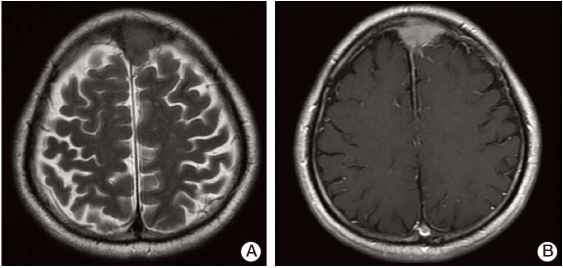
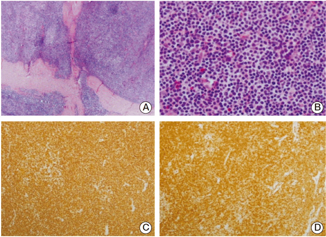
A 69-year-old man visited at our hospital in January 2012 with a 2-month history of headache. The patient had no trauma history and no significant medical history. He had no other objective neurologic finding. A magnetic resonance imaging (MRI) scan of brain showed a relatively homogeneous enhancing 1.9×3.6-cm-sized extra-axial mass with a broad based dural attachment to the anterosuperior aspect of the falx cerebri involving the superior sagittal sinus. The mass showed dural tail sign, and because of suspicious cerebrospinal fluid cleft sign, the mass was considered of dura origin (Fig. 1). He was diagnosed as meningioma radiographically. He underwent bifrontal craniotomy with total removal of the tumor. The mass was completely resected with good dural margins. Pathologic examination revealed that the majority of the tumor cells were monocytoid B-cells, which have small to medium sized nuclei and abundant pale cytoplasm (Fig. 2). There were also remnants of reactive follicles and follicular colonization in the tumor. Immunophenotypically the tumor cells were positive for CD20, bcl-2, and MUM-1 but negative for CD3, CD5, CD10, cyclin D1, and bcl-6. Ki-67 labeling index was 15%. Overall, these features are compatible with extranodal MZBCL of MALT type.
The patient underwent chest computed tomography (CT), abdominal and pelvic CT, spine MRI, bone marrow aspiration and biopsy, and cerebrospinal tapping for staging workups. These did not reveal any abno-rmalities. Ophthalmologic evaluations revealed no other areas of disease involvement. The laboratory finding was normal. Rapid urease test for Helicobacter pylori was negative. After the bifrontal craniotomy with total removal of the tumor, the patient was treated with systemic chemotherapy with cyclophosphamide, vincristine and prednisolone for six cycles without treatment-related complication. Follow-up positron emission tomographic scan showed him to be in disease-free. As of October 2014, our patient has been free of symptoms or recurrence for 2 years and 9 months.
Discussion
MZBCL is a type of B-cell lymphoma presenting primarily in the marginal zone. Extranodal MZBCL of MALT is defined as an extranodal lymphoma, and MALT lymphomas typically present as a local mass without bone marrow and lymph node involvement, in contrast to nodal and splenic MZBCL [5]. The stomach and ocular adnexa are the most common sites for the MALT lymphomas. It is also recognized in intestinal tract, lung, thyroid, salivary glands, skin, liver, CNS, and breast [2].
Primary CNS lymphoma constitutes only 3% of primary brain tumors [6]. The majority of primary CNS lymphoma are diffuse large B-cell lymphoma that are mostly intraparenchymal [7]. T-Cell, low-grade B-cell lymphoma and anaplastic large cell lymphoma of the CNS are extremely rare [7]. Most primary CNS lymphoma presents as a space-occupying lesion within the brain parenchyma and periventricular regions [8]. Primary CNS MZBCL is very rare and indolent and occurs mostly in the extra-axial dura, in contrast to primary CNS high-grade lymphoma [1]. Cases of dural MZBCL in the literature are reviewed in Table 1.
We reported a case of a MALT-type lymphoma of the dura presenting symptomatically and radiographically as meningioma. Primary CNS MZBCL arises mostly from areas rich in meningothelial cells or throughout the arachnoid membrane and arachnoid villi within the dural venous sinus [1]. Because meningiomas are also common to these sites, dural MALT lymphoma has often been misdiagnosed as meningioma. CT or MRI findings reveal well defined, intracranial, dural mass. Presenting symptoms vary by tumor location; patients usually present with headache, meningeal signs, and cranial nerve involvement [8,9].
The diagnosis in our case was confirmed by the histological and immunophenotyping findings, but CNS MALT lymphoma can be difficult to identify even with immunophenotyping. In our case, immunohistochemical stains were negative for CD3, CD5, CD10, while CD20 was positive.
There is no defined treatment guideline for this rare clinical entity. Primary CNS MALT lymphoma originating from the dura matter is curable by surgical removal and radiation therapy compared to parenchymal primary CNS lymphoma or systemic lymphoma with CNS metastasis [9]. Although no standard of care is established, it would seem reasonable to treat with aggressive radiotherapy, with or without surgical resection, depending on the amount of mass [10]. The radiation field is determined by the extent of the dural lesions and leptomeningeal involvement. Therefore, this rare subtype is important to distinguish from other ordinary B-cell NHL with poor clinical outcomes. In some cases, chemotherapy might be suitable as the sole adjuvant treatment. The dura mater is outside the blood-brain barrier, which may explain the increased risk of systemic relapse compare to other types of primary CNS lymphoma. In our case, cyclophosphamide, vincristine and prednisolone chemotherapy brought about a disease-free state.
In summary, we have presented a very rare case of primary CNS MZBCL involving the dura. Longer clinical observation can help to assess the final outcome of patients with primary CNS MALT lymphoma.