Cancer Research and Treatment : Official Journal of Korean Cancer Association 2015;47(2):313-321.
DOI: https://doi.org/10.4143/crt.2013.222
Published online: September 11, 2014
1Department of Respiratory Medicine, The First Affiliated Hospital of Chongqing Medical University, Chongqing, China
2Department of Obstetrics and Gynecology, The Shanxi Da Yi Hospital, Taiyuan, China
Correspondence: Zan-Hong Wang, PhD Department of Obstetrics and Gynecology, The Shanxi Da Yi Hospital, Taiyuan, 030001, China Tel: 86-351-4366608 Fax: 86-351-4366608 E-mail: wangzanhong@126.com
• Received: October 30, 2013 • Accepted: February 21, 2014
This is an Open-Access article distributed under the terms of the Creative Commons Attribution Non-Commercial License (http://creativecommons.org/licenses/by-nc/3.0/) which permits unrestricted non-commercial use, distribution, and reproduction in any medium, provided the original work is properly cited.
Autophagy is one of the ways to degrade unfolded proteins after endoplasmic reticulum (ER) stress. The purpose of this study is to determine whether a blockade of autophagy leads to aggravated endoplasmic reticulum stress, which then induces cells apoptosis in HeLa cells treated with paclitaxel.
Materials and Methods
Autophagy activation and the proapoptotic effects were characterized using monodansylcadaverine labeling and Hoechest staining, respectively. A Western blot analysis was used to detect the expression of apoptotic and autophagy-related genes. A flow cytometry was used to assess the cell apoptosis ratio.
Results
Paclitaxel exposure induced the aggregation of autophagosomes in the cytoplasms of cervical cancer HeLa cells. The expression of Beclin 1 and LC3 II were upregulated, but p62 was downregulated, which suggests that autophagy was promoted by paclitaxel. On the other hand, the expression of GRP78 obviously increased, suggesting that ER stress was induced after paclitaxel treatment. The cell proliferation assay indicated that a knockdown of Beclin 1 sensitized HeLa cells to paclitaxel. Furthermore, paclitaxel-mediated apoptotic cell death was further potentiated by the pretreatment with autophagy inhibitor chloroquine or small interfering RNA against Beclin 1. These results suggest that an induction of autophagy by paclitaxel may induce cell survival rather than cell death in HeLa cells; moreover, inhibition of autophagy led to an aggravated ER stress and an induction of downstream apoptosis.
Conclusion
Our results reveal autophagy induced by paclitaxel conferred protection of tumor cells against apoptosis, and blockade of autophagy subsequently aggravated ER stress, enhancing the apoptosis associated with paclitaxel treatment in HeLa cells.
Paclitaxel is one of the most effective chemotherapeutic agents and is widely used in the treatment of tumors; however, its acquired resistance limits its usage. Recently, several studies have found that paclitaxel can induce autophagy. Eum and Lee [1] reported that paclitaxel induced a notable increase of autophagy in v-Ha-ras-transformed NIH 3T3 cells. In a particular study, promotion of apoptotic cell death by paclitaxel was accompanied with an induction of autophagy in A549 cells. However, the underlying mechanism is still not illustrated.
Various evidences have suggested that autophagy may play dual roles during chemotherapy. Sometimes, autophagy is a protective pathway associated with the maintenance of cellular homeostasis activated in response to nutrient deprivation and various metabolic stressful stimuli. Sometimes, autophagy is a way to induce cancer cell death [2]. Endoplasmic reticulum (ER) stress is a major regulator of autophagy. PERK, IRE1, and increased cytosolic calcium have been implicated as the mediators of ER stress induced autophagy in mammalian cells [3]. ER stress leads to evolutionarily conserved cell stress response, the unfolded protein response (UPR), which induces the expression of chaperones and proteins involved in the recovery process. Moreover, the UPR may upregulate the autophagy machinery [4,5].
ER stress can induce autophagy, and autophagy activation can degrade unfolded aggravated proteins to alleviate ER stress, thus enabling the cell to avoid ER-mediated apoptosis. Therefore, we hypothesized that the inhibition of autophagy may lead to aggravated ER stress, inducing downstream apoptosis. In this study, we tested whether autophagy suppression will exacerbate ER stress and induce ER stress related apoptosis.
Materials and Methods
1. Cell culture, drug treatment, and transfection
The human cervical cancer HeLa cell line was purchased from the Type Culture Collection of the Chinese Academy of Sciences (Shanghai, China). HeLa cells were cultured in Dulbecco's modified Eagle's medium (DMEM) medium that contained 10% fetal bovine serum (Gibco, Germantown, MD), penicillin (10 U/mL, Sigma, St. Louis, MO), and streptomycin (0.1 mg/mL, Sigma), and were kept in a humidified atmosphere of 5% CO2 at 37°C. The cells were exposed to paclitaxel (1 μg/mL, Sigma) or chloroquine (CQ; 5 μM, Sigma) when the confluency reached 50%. Pre-designed siRNAs were purchased for Beclin-1 (Qiagen, Germantown, MD), and one negative random siRNA, exhibiting no significant sequence similarity to human, mouse, or rat gene sequence served as a negative control. The equimolar amounts of siRNAs were incubated with Lipofectamine 2000 Transfection Reagent from Invitrogen (Madison, WI) in accordance to the manufacturer’s instructions.
2. Western blot analysis
The cultured cells were washed twice with phosphate buffered saline (PBS) and lysed with a cold RIPA lysis buffer containing protease inhibitors (phenylmethylsulphonyl fluoride 1 mmol/L and leupeptin 0.1 g/L). Cell lysates were collected from culture plates using a rubber policeman, and proteins were collected by centrifugation. Protein concentrations were determined by bicinchoninic acid protein assay (Pierce, Rockford, IL). Aliquots of 40 μg of proteins were boiled in 2× loading buffer (0.1 M Tris-Cl, pH 6.8, 4% sodium dodecyl sulfate, 0.2% bromophenyl blue, and 20% glycerol) for 10 minutes, loaded into 10% Tris-HCl polyacrylamide gels, and transferred electrophoretically to Immobilon-P membrane (Millipore Corporation, Billerica, MA). The membranes were incubated with primary antibodies and appropriate horseradish peroxidase secondary antibodies. The membranes were additionally probed with an antibody against actin (Santa Cruz Biotechnology, Dallas, TX) to ensure equal loading of proteins between the samples. Detection was performed with chemiluminescent agents (Pierce).
3. Polyclonal antibodies
Anti-beclin 1 antibody (Santa Cruz Biotechnology) recognizes a single 60-kDa band on a Western blot. Anti-LC3 antibody (Santa Cruz Biotechnology) recognizes 16-kDa and 18-kDa two bands. Anti-p62 antibody (Cell Signaling, Danvers, MA), anti–glucose-regulated protein-78 (GRP78) antibody (Cell Signaling), anti-PERK antibody (Cell Signaling), anti-CHOP antibody (Cell Signaling), and anti-IRE1 antibody (Cell Signaling) recognize a single 62-kDa, 78-kDa, 140-kDa, 27-kDa, and 130-kDa band on Western blot, respectively.
4. Cell proliferation assay
HeLa cells were plated in 96-well plates at 5,000 or 6,000 cells per well, respectively. On the following day, the cells were treated with or without 1 μg/mL paclitaxel with five to six replicates for 24 hours or 48 hours. According to the experiment, the cytotoxity was assessed by incubating cells with WST-1 reagent (Beyotime, Beijing, China) for 2 hours, measuring the absorbance at 450 nm, and at 630 nm as reference, with a microplate reader (Bio-Rad, Hercules, CA). The cell survival rate was calculated as follows: (ODexperimental group– ODcontrol group)/ODcontrol group×100%.
5. Assessment of cell apoptosis by flow cytometry
Apoptosis was quantified by a combined staining of annexin V and propidium iodide (PI), using AnnexinV-FITC Apoptosis Detection Kit (Beyotime). In brief, the cells were harvested and resuspended in a 500 μL of 1× binding buffer, after treatment with paclitaxel (1 μg/mL) for 24 hours or paclitaxel treatment followed by CQ (5 μM) treatment for 48 hours. After adding 5 μL of Annexin V-FITC solution and 5 KL of PI solution, the cells were incubated for 15 minutes at room temperature in the dark. At the end of incubation, the cells were analyzed on a FACScan flow cytometry system (Becton Dickinson, San Joes, CA). Data were analyzed using a winMDIv2.8 software (Perdue University Cytometry Laboratories, West Lafayette, IN). All experiments were performed three times for each experimental condition.
6. Hoechest 33258 staining assay
Replicate cultures of 1×10⁶ cells per well were plated in a 24-well plate. The cells were transfected with siRNA and/or treated with paclitaxel. After a change of the fresh medium 24 hours later, the cells were incubated with 5 μL of Hoechst 33258 (Beyotime) solution per well at 37°C for 10 minutes, followed by observation under a fluorescence microscope. Strong fluorescence can be observed in the nuclei of apoptotic cells, while weak fluorescence was observed in non-apoptotic cells. Quantification of apoptotic cells was performed via taking the images in random fields and counting at least 200 cells in four random fields in each well.
7. Quantification of MDC labeling of cells
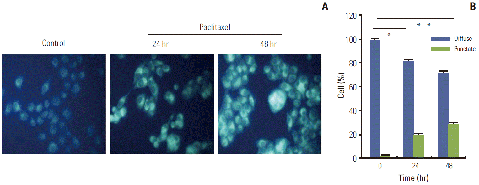
The autofluorescent agent monodansylcadaverine (MDC; Sigma) was recently introduced as a specific autophagolysosome marker to analyze the autophagic process. HeLa cells were treated with paclitaxel 1 μg/mL for 24 hours. Autophagic vacuoles were labeled with MDC by incubating the cells with 0.05 mM MDC in DMEM at 37°C for 10 minutes, at various times after the paclitaxel treatment. After incubation, the cells were washed three times with PBS and immediately analyzed with a fluorescence microscope (Nikon Eclipse TE 300, Nikon, Tokyo, Japan) equipped with a filter system (V-2A excitation filter, 380/420 nm; barrier filter, 450 nm). Images were captured with a CCD camera and imported into Adobe Photoshop.
8. Statistical analysis
Statistical analysis of the results was performed by analysis of variance (ANOVA) and Tukey multiple range tests, using a Prism ver. 2.0 software (GraphPad Software, San Diego, CA). Statistical significance was defined as p ≤ 0.05.
Results
1. Autophagy was induced in HeLa cells treated with paclitaxel
An increasing number of studies have shown that autophagy is activated during stress conditions for the degradation of superfluous proteins and damaged organelles. In line with these studies, we observed that (as shown in Fig. 1), compared with the control cells, HeLa cells treated with paclitaxel showed an increase in the number of MDC labeled vesicles, suggesting an induction of autophagic vacuoles formation after paclitaxel treatment. Furthermore, autophagy related genes were up-regulated. Autophagy marker gene Beclin 1 and LC3 II were induced; however, p62 was downregulated by paclitaxel treatment (Fig. 2). The results indicated that autophagy was induced by paclitaxel exposure in HeLa cells.
2. Paclitaxel induced ER stress in HeLa cells
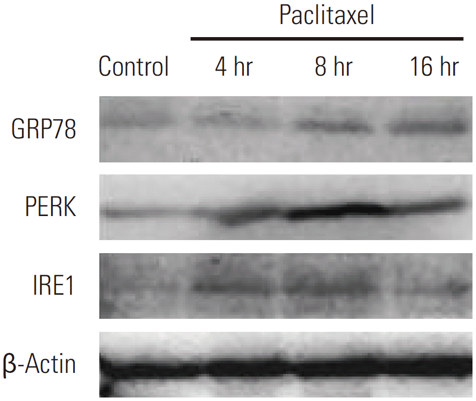
Recent literature suggests that disturbances in the integrity of ER activate autophagy [6]. To investigate the molecular mechanisms behind paclitaxel induced autophagy in HeLa cells, we investigated whether paclitaxel can trigger ER stress. To address this issue, HeLa cells treated with paclitaxel were detected by Western blotting. GRP78 is an ER chaperone protein, which is upregulated by ER stress. When the cells were treated with paclitaxel, GRP78 protein expression was significantly increased at 8 hours and 16 hours (Fig. 3). Our data further showed that paclitaxel induced ER stress was accompanied by the upregulation of PERK and IRE1 in HeLa cells. Both PERK and IRE1 are regulated by GRP78 during ER stress (Fig. 3). These observations indicated that paclitaxel can induce ER stress in HeLa cells.
3. Blocking autophagy exacerbated UPR
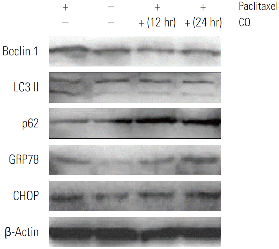
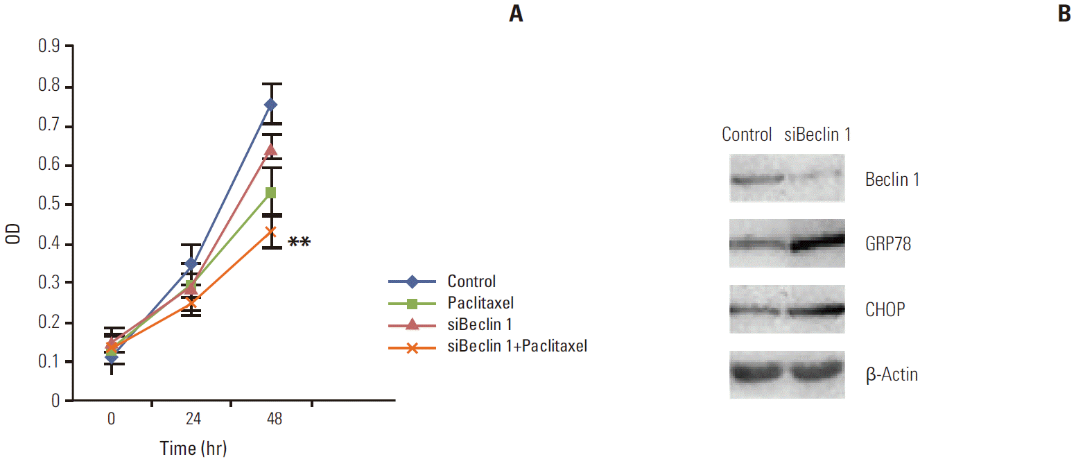
Since we observed that HeLa cells exposed to paclitaxel undergo both ER stress and autophagy, we hypothesized that autophagy in cervical cancer cells is activated by paclitaxel through an induction of ER stress. Firstly, we used autophagy pharmaceutical inhibitor CQ to treat HeLa cells after paclitaxel exposure to detect UPR related genes expression. Our results show that CQ administration, following paclitaxel treatment, predominantly induced the expression of p62 and blocked the increase in the expression of LC3B and Beclin 1, while elevating the expression of GRP78 and CHOP (as shown in Fig. 4). To firmly establish the effects of autophagy inhibition on ER stress in HeLa cells after paclitaxel treatment, Beclin 1 was transiently silenced using siRNA. Our results showed that the cytotoxic effect of paclitaxel was significantly increased by blocking Beclin 1 expression when compared to the random control (Appendix 1A). To gain further insight into this mechanism, we detected the expression of GRP78 and CHOP (Appendix 1A and B). As expected, the expression of GRP78 and CHOP were enhanced. These results support our hypothesis that paclitaxel could induce accumulation of unfolded or misfolded proteins, while autophagy could clear them to attenuate ER stress in HeLa cells.
4. Inhibition of autophagy potentiates ER stress-mediated apoptotic cell death
Accumulation of unfolded or misfolded proteins in the ER lumen induced activation of chaperones and the recovery process. When the recovery process autophagy was inhibited, severe ER stress causes cell death, usually by an activation of intrinsic apoptosis. Therefore, we next observed the cell apoptosis ratio and tested the expression of ER stress related apoptosis genes after autophagy inhibition. As shown in Fig. 5, cell apoptosis induction by paclitaxel was markedly enhanced in the presence of CQ. Consistent with the results using CQ, the degree of apoptosis induced by paclitaxel was also enhanced when Beclin 1 protein level was reduced by a specific siRNA, as observed with Hoechest 33258 staining (Fig. 6). More apoptotic cells were observed in Beclin 1 siRNA–transfected cells after paclitaxel treatment than in the control cells treated with paclitaxel. These results demonstrate that autophagy induced by paclitaxel conferred protection of tumor cells against apoptosis, and blockade of autophagy subsequently enhanced the apoptosis associated with paclitaxel treatment.
Discussion
Cervical cancer is the most common malignancy of the female reproductive system. Although neoadjuvant chemotherapy, along with concurrent chemo- and radiotherapies have benefited majority patients, survival in women with recurrent or metastatic cervical cancer remains poor. Resistance of cancer to chemotherapy is one of the primary causes of treatment failure [6,7]. Paclitaxel is the prototypic member of the taxane class of mitotic inhibitor-based chemotherapies. Paclitaxel and its analogs are widely used in the treatment of solid tumors, including cervical cancer, especially in a combination with platinum [8]. Therefore, other therapeutic approaches must be tested in order to further improve the prognosis.
Many studied have shown that antitumor activity of paclitaxel is due to its interaction with b-tubulin, where it inhibits microtubule depolymerization and arrests cycling cells in the mitotic (M) phase. Finally, paclitaxel activates the apoptotic signal pathway, leading to caspase activation, which involves the Bcl-2 family of proteins and the mitochondria [9,10]. Recently, the ER was reported to be a cytosolic target of paclitaxel induced apoptosis via the ER stress pathway; sustained ER stress induces caspase-mediated apoptosis [11]. However, the exact mechanisms of paclitaxelinduced cell death are not completely illustrated.
ER is a central cell organelle that is involved in biosynthesis, protein folding, and modification of proteins [12]. ER is very sensitive to minor perturbations in its environment. A number of cellular stress conditions, such as glucose deprivation, oxidative stress, and infection, can lead to the accumulation of unfolded or misfolded proteins in the ER lumen, which disturbs the ER function and result in ER stress [13]. ER stress leads to the UPR to avoid cell damage, which triggers two downstream signaling events. One is the induction of genes encoding for ER resident chaperones to properly fold the unfolded proteins and the other being suppression of initiation of protein synthesis [14]. However, severe ER stress can cause cell death, usually by an activation of apoptosis [15]. Moreover, in order to clear the ER of accumulated terminally misfolded protein aggregates that cannot be degraded by the proteasome, the UPR may upregulate the autophagy machinery [16,17].
Autophagy plays an important physiological role in the turnover of cellular proteins and other macromolecules. Autophagy can be induced by a change in the environmental conditions, such as nutrient depletion; moreover, autophagy is involved in development, differentiation, and tissue remodeling in various organisms [18,19]. In addition, autophagy can also be induced in a response to chemotherapy and has been considered as an important mechanism contributing to drug resistance, and the autophagic process may protect cancer cells from toxicity [20]. Therefore, the inhibition of autophagy may result in a greater susceptibility to cell death-inducing mechanisms.
In our study, we hypothesized that inhibition of autophagy leads to aggravated ER stress, which then induces downstream apoptosis. We firstly detected the expression of autophagy marker genes Beclin 1, LC3II, and p62 in HeLa cells treatment with paclitaxel, and then detected the expression of ER stress marker genes GRP78 and CHOP. Although the results showed that the expression of these genes had been changed after paclitaxel treatment, a conclusion cannot be made since autophagy and ER stress are closely related. Then, we detected the expression of ER stress related genes in HeLa cells after the treatment with an inhibitor of autophagy or siRNA against the autophagy gene to confirm the relationship between autophagy and ER stress. The results demonstrated that a blockade of autophagy can increase the expression of the ER stress genes. Moreover, ER stress-mediated apoptotic cell death after paclitaxel treatment was potentiated by an inhibition of autophagy. These results preliminarily indicated that inhibition of autophagy led to the aggravation of ER stress and potentiated downstream cell apoptosis. CQ is a lysosomotropic drug that raises intralysosomal pH level and impairs autophagic protein degradation [21]. By blocking the last step of the autophagy pathway, CQ can increase protein accumulation in the cells more than 3-methyladenine (3MA) which is known to be a representative autophagy inhibitor by mainly blocking the initiation of autophagy pathway. To test our hypothesis, we used CQ but not 3MA to treat HeLa cells to induce protein accumulation; then detected the expression of the ER stress genes and cell apoptosis. The result showed that p62 was dramatically elevated after CQ treatment, which means protein degradation was significantly blocked and more protein accumulated. We also wanted to know whether the inhibition of the initiation of autophagy could aggravate ER stress as such, we used siRNA against Beclin 1 to test this. The results showed that either CQ or siRNA against Beclin 1 could promote the expression of ER stress genes and potentiate downstream cell apoptosis. Therefore, a blockade of autophagy could aggravate ER stress and promote cell apoptosis in HeLa cells.
Xi et al. [22] reported that paclitaxel can promote apoptotic cell death, accompanied with an induction of autophagy in lung cancer A549 cells. Kim et al. [23] also revealed that paclitaxel can increase autophagic activity, suppressing the autophagy enhancement of paclitaxel-induced apoptosis in OS cells. Conversely, the over-expression of Beclin1 in cervical cancer CaSki cells may enhance apoptosis signaling induced by anti-cancer drugs (cisplatin, paclitaxel, 5-fluorouracil, and epirubicin), which suggests that autophagy can promote apoptosis after drug treatment [7]. In mitotic cells, paclitaxel can block the activation of class III phosphatidyl inositol 3 kinase, Vps34, a critical initiator of autophagosome formation. Chemically or genetically blocking autophagosome formation diminished paclitaxel-induced cell death, suggesting that autophagosome accumulation sensitized the cells to paclitaxel toxicity [8]. Interestingly, in v-Ha-ras-transformed NIH 3T3 cells treated with paclitaxel, both autophagy and apoptosis act as cooperative partners to induce cell death [1]. Paclitaxel treatment induced cell death through autophagy or apoptosis or both is different in different cells. Furthermore, autophagy sometimes leads to cell death, while other times protects cells from cell death. In our study, protective autophagy was induced after paclitaxel treatment in HeLa cells.
In U937 cells, ER would seem to contribute to paclitaxelinduced apoptosis via both the early release of Ca2+ and the late amplification of mitochondria-mediated apoptotic signals [24]. Thapsigargin, a classic ER stress inducer, can increase the cell killing potency of paclitaxel or docetaxel by 10- to 12-fold in Human prostate cancer PC-3 cells. In addition, both the intrinsic and extrinsic apoptotic pathways were involved in the sensitization mechanisms. ER stress-apoptotic response is conducive to chemotherapeutic sensitization [25]. Our research reveals that the inhibition of autophagy can intensify ER stress apoptotic response and sensitivity to paclitaxel.
Conclusion
Our results preliminarily showed that the inhibition of autophagy can intensify ER stress and induce downstream cell apoptosis in HeLa cells. The main finding shown in our study is consistent with previous studies, but our results contribute new knowledge to understand the role of ER-stress related autophagy in cancer cells after taxane treatment.
NOTES
Conflict of interest relevant to this article was not reported.
Acknowledgements
The research is supported by Key Science and Technology Program of Shanxi Province (Grant No. 20120313019-2), Foundation of Public Health Department of Shanxi (Grant No. 2011024).
Fig. 1.
Monodansylcadaverine (MDC) labeled vesicles in HeLa cells treated with paclitaxel. HeLa cells were treated with paclitaxel 1 μg/mL for 24 hours. Autophagic vacuoles were labeled with MDC by incubating cells with 0.05 mM MDC in Dulbecco’s modified Eagle’s medium at 37°C for 10 minutes at various times after paclitaxel treatment. Compared with the control cells, HeLa cells treated with paclitaxel showed an increase in the number of MDC labeled vesicles, suggesting an induction of autophagic vacuoles formation after paclitaxel treatment. (A) The image of MDC labeled vesicles in HeLa cells. (B) The quantitative result of MDC labeled vesicles. *p < 0.05, **p < 0.01.
Fig. 2.
Detection of autophagy related genes expression by Western blotting in HeLa cells after treatment with paclitaxel for 24 hours and 48 hours. HeLa cells were treated with paclitaxel 1 μg/mL for 24 hours and 48 hours and then cell lysates were collected. Autophagy marker gene Beclin 1 and LC3 II were induced, but p62 was downregulated by paclitaxel treatment, indicating that autophagy was induced by paclitaxel exposure in HeLa cells.
Fig. 3.
Detection of endoplasmic reticulum stress related genes expression by Western blotting in HeLa cells after treatment with paclitaxel. HeLa cells were treated with paclitaxel 1 μg/mL for 4 hours, 8 hours, and 16 hours and then cell lysates were collected. Glucose-regulated protein-78 (GRP78) protein expression was significantly increased at 8 hours and 16 hours, and the upregulation of PERK and IRE1 in HeLa cells were observed.
Fig. 4.
Detection of autophagy and endoplasmic reticulum stress related genes expression by Western blotting in HeLa cells after treatment with autophagy inhibitor chloroquine (CQ) for 12 hours and 24 hours following paclitaxel exposure. Firstly, HeLa cells were treated with paclitaxel 1 μg/mL for 2 hours, then treated with 5 μM CQ for 12 hours and 24 hours. CQ administration following paclitaxel treatment predominantly induced expression of p62, blocked the increase in the expression of LC3 II and Beclin 1, and meanwhile elevated the expression of glucose-regulated protein-78 (GRP78) and CHOP.
Fig. 5.
Apoptosis ratio of HeLa cells treated with paclitaxel in the presence of chloroquine (CQ). HeLa cells were harvested and resuspended in binding buffer, after treatment with paclitaxel (1 μg/mL) for 24 hours or paclitaxel treatment followed by CQ (5 μM) treatment for 48 hours. Cell apoptosis were then analyzed by flow cytometry. The percentage of cells with a sub-G1 DNA content was taken as a measure of cell death. At least three independent experiments were performed. *p < 0.05, **p < 0.01. Cell apoptosis induction by paclitaxel was markedly enhanced in the presence of CQ. PI, propidium iodide; PCX, paclitaxel.
Fig. 6.
Hoechest 33258 staining of HeLa cells after different treatment. The cells were transfected with siRNA and/or treated with paclitaxel (1 μg/mL) for 24 hours. After a change of fresh medium, the cells were incubated with Hoechst 33258 solution followed by observation under a fluorescence microscope. Strong fluorescence can be observed in the nuclei of apoptotic cells, while weak fluorescence was observed in non-apoptotic cells. Quantification of apoptotic cells was performed by taking the images in random fields and counting at least 200 cells in four random fields in each well. More apoptotic cells were observed in Beclin 1 siRNA–transfected cells after paclitaxel treatment than in control cells treated with paclitaxel. PCX, paclitaxel. *p < 0.05, **p < 0.01.
REFERENCES
1. Eum KH, Lee M. Crosstalk between autophagy and apoptosis in the regulation of paclitaxel-induced cell death in v-Haras-transformed fibroblasts. Mol Cell Biochem. 2011;348:61–8. Article
2. Levine B, Sinha S, Kroemer G. Bcl-2 family members: dual regulators of apoptosis and autophagy. Autophagy. 2008;4:600–6. ArticlePubMedPMC
3. Kouroku Y, Fujita E, Tanida I, Ueno T, Isoai A, Kumagai H, et al. ER stress (PERK/eIF2alpha phosphorylation) mediates the polyglutamine-induced LC3 conversion, an essential step for autophagy formation. Cell Death Differ. 2007;14:230–9. ArticlePubMed
4. Ogata M, Hino S, Saito A, Morikawa K, Kondo S, Kanemoto S, et al. Autophagy is activated for cell survival after endoplasmic reticulum stress. Mol Cell Biol. 2006;26:9220–31. ArticlePubMedPMC
5. Xu Y, Yu H, Qin H, Kang J, Yu C, Zhong J, et al. Inhibition of autophagy enhances cisplatin cytotoxicity through endoplasmic reticulum stress in human cervical cancer cells. Cancer Lett. 2012;314:232–43. ArticlePubMed
6. Suh DH, Kim JW, Kim K, Kim HJ, Lee KH. Major clinical research advances in gynecologic cancer in 2012. J Gynecol Oncol. 2013;24:66–82. ArticlePubMedPMC
7. Sun Y, Liu JH, Jin L, Lin SM, Yang Y, Sui YX, et al. Overexpression of the Beclin1 gene upregulates chemosensitivity to anti-cancer drugs by enhancing therapy-induced apoptosis in cervix squamous carcinoma CaSki cells. Cancer Lett. 2010;294:204–10. ArticlePubMed
8. Veldhoen RA, Banman SL, Hemmerling DR, Odsen R, Simmen T, Simmonds AJ, et al. The chemotherapeutic agent paclitaxel inhibits autophagy through two distinct mechanisms that regulate apoptosis. Oncogene. 2013;32:736–46. ArticlePubMed
9. Jordan MA, Toso RJ, Thrower D, Wilson L. Mechanism of mitotic block and inhibition of cell proliferation by taxol at low concentrations. Proc Natl Acad Sci U S A. 1993;90:9552–6. ArticlePubMedPMC
10. Shi J, Orth JD, Mitchison T. Cell type variation in responses to antimitotic drugs that target microtubules and kinesin-5. Cancer Res. 2008;68:3269–76. ArticlePubMed
11. Gorka M, Daniewski WM, Gajkowska B, Lusakowska E, Godlewski MM, Motyl T. Autophagy is the dominant type of programmed cell death in breast cancer MCF-7 cells exposed to AGS 115 and EFDAC, new sesquiterpene analogs of paclitaxel. Anticancer Drugs. 2005;16:777–88. ArticlePubMed
12. Gorlach A, Klappa P, Kietzmann T. The endoplasmic reticulum: folding, calcium homeostasis, signaling, and redox control. Antioxid Redox Signal. 2006;8:1391–418. ArticlePubMed
13. Kim I, Xu W, Reed JC. Cell death and endoplasmic reticulum stress: disease relevance and therapeutic opportunities. Nat Rev Drug Discov. 2008;7:1013–30. ArticlePubMed
14. Kandala PK, Srivastava SK. Regulation of macroautophagy in ovarian cancer cells in vitro and in vivo by controlling glucose regulatory protein 78 and AMPK. Oncotarget. 2012;3:435–49. ArticlePubMedPMC
15. Szegezdi E, Logue SE, Gorman AM, Samali A. Mediators of endoplasmic reticulum stress-induced apoptosis. EMBO Rep. 2006;7:880–5. ArticlePubMedPMC
16. Kroemer G, Marino G, Levine B. Autophagy and the integrated stress response. Mol Cell. 2010;40:280–93. ArticlePubMedPMC
17. Hoyer-Hansen M, Jaattela M. Connecting endoplasmic reticulum stress to autophagy by unfolded protein response and calcium. Cell Death Differ. 2007;14:1576–82. ArticlePubMed
18. Boya P, Gonzalez-Polo RA, Casares N, Perfettini JL, Dessen P, Larochette N, et al. Inhibition of macroautophagy triggers apoptosis. Mol Cell Biol. 2005;25:1025–40. ArticlePubMedPMC
19. Sridhar S, Botbol Y, Macian F, Cuervo AM. Autophagy and disease: always two sides to a problem. J Pathol. 2012;226:255–73. ArticlePubMedPMC
21. Amaravadi RK, Yu D, Lum JJ, Bui T, Christophorou MA, Evan GI, et al. Autophagy inhibition enhances therapy-induced apoptosis in a Myc-induced model of lymphoma. J Clin Invest. 2007;117:326–36. ArticlePubMedPMC
22. Xi G, Hu X, Wu B, Jiang H, Young CY, Pang Y, et al. Autophagy inhibition promotes paclitaxel-induced apoptosis in cancer cells. Cancer Lett. 2011;307:141–8. ArticlePubMed
23. Kim HJ, Lee SG, Kim YJ, Park JE, Lee KY, Yoo YH, et al. Cytoprotective role of autophagy during paclitaxel-induced apoptosis in Saos-2 osteosarcoma cells. Int J Oncol. 2013;42:1985–92. ArticlePubMed
24. Liao PC, Tan SK, Lieu CH, Jung HK. Involvement of endoplasmic reticulum in paclitaxel-induced apoptosis. J Cell Biochem. 2008;104:1509–23. ArticlePubMed
25. Wu Y, Fabritius M, Ip C. Chemotherapeutic sensitization by endoplasmic reticulum stress: increasing the efficacy of taxane against prostate cancer. Cancer Biol Ther. 2009;8:146–52. ArticlePubMedPMC
Appendix
Appendix 1.
The effect of autophagy inhibition on endoplasmic reticulum stress. Firstly, HeLa cells were treated with paclitaxel 1 μg/mL for 2 hours, then transiently transfected with siRNA for another 24-hour and 48-hour culture. (A) Result of cell proliferation assay. The cytotoxic effect of paclitaxel was significantly increased by blocking Beclin 1 expression when compared to the control. (B) Result of Western blotting. The expression of glucose-regulated protein-78 (GRP78) and CHOP were enhanced when Beclin 1 expression was knock down.
Figure & Data
REFERENCES
Citations
Citations to this article as recorded by
Endoplasmic reticulum stress induced autophagy in cancer and its potential interactions with apoptosis and ferroptosis Haitang Liao, Shuang Liu, Qiang Ma, He Huang, Arul Goel, Pedram Torabian, Chakrabhavi Dhananjaya Mohan, Chenyang Duan Biochimica et Biophysica Acta (BBA) - Molecular Cell Research.2025; 1872(1): 119869. CrossRef
Autophagy in cancer resistance to paclitaxel: Development of combination strategies Jan Škubník, Vladimíra Svobodová Pavlíčková, Tomáš Ruml, Silvie Rimpelová Biomedicine & Pharmacotherapy.2023; 161: 114458. CrossRef
Advances in autophagy modulation of natural products in cervical cancer Tao Tao, Ping Zhang, Zhi Zeng, Min Wang Journal of Ethnopharmacology.2023; 314: 116575. CrossRef
Chloroquine Sensitizes Esophageal Carcinoma EC109 Cells to Paclitaxel by Inhibiting Autophagy
Zichun Yuan, Jiajing Cai, Qin Du, Qiang Ma, Lei Xu, Yan Cai, Xiaowu Zhong, Xiaolan Guo Critical Reviews in Eukaryotic Gene Expression.2023; 33(6): 43. CrossRef
New sight into interaction between endoplasmic reticulum stress and autophagy induced by vanadium in duck renal tubule epithelial cells Li Wang, Yueying Pan, Fan Yang, Xiaowang Guo, Junjun Peng, Xiaoyu Wang, Yukun Fang, Jing Chen, Xin Yi, Huabin Cao, Guoliang Hu Chemico-Biological Interactions.2022; 362: 109981. CrossRef
Cisplatin and paclitaxel-loaded liposomes induced cervical cancer (HeLa) cell death with multiple copies of human papillomavirus by apoptosis and decreased their cytotoxic effect on non-tumor cells Paulo Emilio Feuser, Ellen De Pieri, Maria Eduarda Oliveira, Arthur Poester Cordeiro, Rodrigo Cercena, Pedro Henrique Hermes de Araújo, Alexandre Gonçalves Dal Bó, Ricardo Andrez Machado-de-Ávila Journal of Drug Delivery Science and Technology.2022; 73: 103457. CrossRef
Mechanisms of cancer cell death induction by paclitaxel: an updated review Shuang Zhao, Yufei Tang, Ruohan Wang, Masoud Najafi Apoptosis.2022; 27(9-10): 647. CrossRef
An Updated Review on Implications of Autophagy and Apoptosis in Tumorigenesis: Possible Alterations in Autophagy through Engineered Nanomaterials and Their Importance in Cancer Therapy Habib Ghaznavi, Milad Shirvaliloo, Amir Zarebkohan, Zinat Shams, Fatemeh Radnia, Zahra Bahmanpour, Saman Sargazi, Ramin Saravani, Sakine Shirvalilou, Omolbanin Shahraki, Sheida Shahraki, Ziba Nazarlou, Roghayeh Sheervalilou Molecular Pharmacology.2021; 100(2): 119. CrossRef
Demethylzeylasteral (T-96) initiates extrinsic apoptosis against prostate cancer cells by inducing ROS-mediated ER stress and suppressing autophagic flux Dong-lin Yang, Ya-jun Zhang, Liu-jun He, Chun-sheng Hu, Li-xia Gao, Jiu-hong Huang, Yan Tang, Jie Luo, Dian-yong Tang, Zhong-zhu Chen Biological Research.2021;[Epub] CrossRef
Perturbation-Based Modeling Unveils the Autophagic Modulation of Chemosensitivity and Immunogenicity in Breast Cancer Cells Isaac Quiros-Fernandez, Lucía Figueroa-Protti, Jorge L. Arias-Arias, Norman Brenes-Cordero, Francisco Siles, Javier Mora, Rodrigo Antonio Mora-Rodríguez Metabolites.2021; 11(9): 637. CrossRef
Enhanced efficacy of propranolol therapy for infantile hemangiomas based on a mesoporous silica nanoplatform through mediating autophagy dysfunction Haiwei Wu, Xuan Wang, Hao Liang, Jiawei Zheng, Shengyun Huang, Dongsheng Zhang Acta Biomaterialia.2020; 107: 272. CrossRef
Loss of tumor susceptibility gene 101 (TSG101) perturbs endoplasmic reticulum structure and function Zenia Kaul, Debdatto Mookherjee, Subhrangshu Das, Debmita Chatterjee, Saikat Chakrabarti, Oishee Chakrabarti Biochimica et Biophysica Acta (BBA) - Molecular Cell Research.2020; 1867(9): 118741. CrossRef
RSK2 protects human breast cancer cells under endoplasmic reticulum stress through activating AMPKα2-mediated autophagy Lan-Ya Li, Xi-Sha Chen, Kuan-Song Wang, Yi-Di Guan, Xing-Cong Ren, Dong-Sheng Cao, Xin-Yuan Sun, Ao-Xue Li, Yong-Guang Tao, Yi Zhang, Ming-Zhu Yin, Xin-Luan Wang, Ming-Hua Wu, Jin-Ming Yang, Yan Cheng Oncogene.2020; 39(43): 6704. CrossRef
New Insights into Therapy-Induced Progression of Cancer Polina V. Shnaider, Olga M. Ivanova, Irina K. Malyants, Ksenia S. Anufrieva, Ilya A. Semenov, Marat S. Pavlyukov, Maria A. Lagarkova, Vadim M. Govorun, Victoria O. Shender International Journal of Molecular Sciences.2020; 21(21): 7872. CrossRef
Kaempferol Induces Cell Death in A2780 Ovarian Cancer Cells and Increases Their Sensitivity to Cisplatin by Activation of Cytotoxic Endoplasmic Reticulum-Mediated Autophagy and Inhibition of Protein Kinase B Attalla Farag El-Kott, A. A. Shati, M. A. Al-Kahtani, S. A. Alharbi Folia Biologica.2020; 66(1): 36. CrossRef
Targeted Codelivery of Docetaxel and Atg7 siRNA for Autophagy Inhibition and Pancreatic Cancer Treatment Miaozun Zhang, Wei Zhang, Guping Tang, Hebin Wang, Min Wu, Weiming Yu, Zhenfeng Zhou, Yiping Mou, Xingang Liu ACS Applied Bio Materials.2019; 2(3): 1168. CrossRef
An ATG5 knockout promotes paclitaxel resistance in v-Ha-ras-transformed NIH 3T3 cells Seong Yun Eom, Sung-Hee Hwang, Hojin Yeom, Michael Lee Biochemical and Biophysical Research Communications.2019; 513(1): 234. CrossRef
Resveratrol, an activator of SIRT1, improves ER stress by increasing clusterin expression in HepG2 cells Jinmi Lee, Seok-Woo Hong, Hyemi Kwon, Se Eun Park, Eun-Jung Rhee, Cheol-Young Park, Ki-Won Oh, Sung-Woo Park, Won-Young Lee Cell Stress and Chaperones.2019; 24(4): 825. CrossRef
Molecular Mechanisms Underlying Autophagy-Mediated Treatment Resistance in Cancer Cally J. Ho, Sharon M. Gorski Cancers.2019; 11(11): 1775. CrossRef
A promising natural product, pristimerin, results in cytotoxicity against breast cancer stem cells in vitro and xenografts in vivo through apoptosis and an incomplete autopaghy in breast cancer Buse Cevatemre, Merve Erkısa, Nazlihan Aztopal, Didem Karakas, Pınar Alper, Chrisiida Tsimplouli, Evangelia Sereti, Konstantinos Dimas, Elif I. Ikitimur Armutak, Ebru Gurel Gurevin, Ayca Uvez, Mattia Mori, Simone Berardozzi, Cinzia Ingallina, Ilaria D’Acq Pharmacological Research.2018; 129: 500. CrossRef
Chitosan-based zinc oxide nanoparticle for enhanced anticancer effect in cervical cancer: A physicochemical and biological perspective Henghui Wu, Juxin Zhang Saudi Pharmaceutical Journal.2018; 26(2): 205. CrossRef
Pro-survival autophagy and cancer cell resistance to therapy Chandan Kanta Das, Mahitosh Mandal, Donat Kögel Cancer and Metastasis Reviews.2018; 37(4): 749. CrossRef
The Superior Antitumor Effect of Self-Assembled Paclitaxel Nanofilaments for Lung Cancer Cells Mengyu He, Jiali Zhu, Na Yu, Hui Kong, Xiaoning Zeng, Weiping Xie, Huae Xu Current Drug Delivery.2018; 16(2): 171. CrossRef
Triptolide induces protective autophagy and apoptosis in human cervical cancer cells by downregulating Akt/mTOR activation Guangyi Qin, Ping Li, Zhuowei Xue Oncology Letters.2018;[Epub] CrossRef
Upstream transcription factor 1 prompts malignancies of cervical cancer primarily by transcriptionally activating p65 expression Wen Wang, Shujuan Yao, Hongjing Jiang, Jing Dong, Xiujuan Cui, Xiangyu Tian, Yanyan Guo, Shiqian Zhang Experimental and Therapeutic Medicine.2018;[Epub] CrossRef
Autophagy inhibitors chloroquine and LY294002 enhance temozolomide cytotoxicity on cutaneous melanoma cell lines in vitro Oxana O. Ryabaya, Andrey N. Inshakov, Angelina V. Egorova, Marina A. Emelyanova, Tatiana V. Nasedkina, Alexander S. Zasedatelev, Dmitry A. Khochenkov, Evgenia V. Stepanova Anti-Cancer Drugs.2017; 28(3): 307. CrossRef
Endoplasmic Reticulum Stress and Homeostasis in Reproductive Physiology and Pathology Elif Guzel, Sefa Arlier, Ozlem Guzeloglu-Kayisli, Mehmet Tabak, Tugba Ekiz, Nihan Semerci, Kellie Larsen, Frederick Schatz, Charles Lockwood, Umit Kayisli International Journal of Molecular Sciences.2017; 18(4): 792. CrossRef
Sensitivity to antitubulin chemotherapeutics is potentiated by a photoactivable nanoliposome Xiaobing Wang, Xiufang Liu, Yixiang Li, Pan Wang, Xiaolan Feng, Quanhong Liu, Fei Yan, Hairong Zheng Biomaterials.2017; 141: 50. CrossRef
ESCRT-0 dysfunction compromises autophagic degradation of protein aggregates and facilitates ER stress-mediated neurodegeneration via apoptotic and necroptotic pathways Ryuji Oshima, Takafumi Hasegawa, Keiichi Tamai, Naoto Sugeno, Shun Yoshida, Junpei Kobayashi, Akio Kikuchi, Toru Baba, Akira Futatsugi, Ikuro Sato, Kennichi Satoh, Atsushi Takeda, Masashi Aoki, Nobuyuki Tanaka Scientific Reports.2016;[Epub] CrossRef
The effect of quercetin nanoparticle on cervical cancer progression by inducing apoptosis, autophagy and anti-proliferation via JAK2 suppression Cheng-lin Luo, Yu-qiong Liu, Peng Wang, Chun-hua Song, Kai-juan Wang, Li-ping Dai, Jian-ying Zhang, Hua Ye Biomedicine & Pharmacotherapy.2016; 82: 595. CrossRef
Role of autophagy and lysosomal drug sequestration in acquired resistance to doxorubicin in MCF-7 cells Baoqing Guo, Adam Tam, Stacey A. Santi, Amadeo M. Parissenti BMC Cancer.2016;[Epub] CrossRef
Blocking autophagy enhances meloxicam lethality to hepatocellular carcinoma by promotion of endoplasmic reticulum stress Jingtao Zhong, Xiaofeng Dong, Peng Xiu, Fuhai Wang, Ju Liu, Honglong Wei, Zongzhen Xu, Feng Liu, Tao Li, Jie Li Cell Proliferation.2015; 48(6): 691. CrossRef
8-p-Hdroxybenzoyl Tovarol Induces Paraptosis Like Cell Death and Protective Autophagy in Human Cervical Cancer HeLa Cells Cui Zhang, Yingnan Jiang, Jin Zhang, Jian Huang, Jinhui Wang International Journal of Molecular Sciences.2015; 16(7): 14979. CrossRef
Blockade of Autophagy Aggravates Endoplasmic Reticulum Stress and Improves Paclitaxel Cytotoxicity in Human Cervical Cancer Cells
Fig. 1. Monodansylcadaverine (MDC) labeled vesicles in HeLa cells treated with paclitaxel. HeLa cells were treated with paclitaxel 1 μg/mL for 24 hours. Autophagic vacuoles were labeled with MDC by incubating cells with 0.05 mM MDC in Dulbecco’s modified Eagle’s medium at 37°C for 10 minutes at various times after paclitaxel treatment. Compared with the control cells, HeLa cells treated with paclitaxel showed an increase in the number of MDC labeled vesicles, suggesting an induction of autophagic vacuoles formation after paclitaxel treatment. (A) The image of MDC labeled vesicles in HeLa cells. (B) The quantitative result of MDC labeled vesicles. *p < 0.05, **p < 0.01.
Fig. 2. Detection of autophagy related genes expression by Western blotting in HeLa cells after treatment with paclitaxel for 24 hours and 48 hours. HeLa cells were treated with paclitaxel 1 μg/mL for 24 hours and 48 hours and then cell lysates were collected. Autophagy marker gene Beclin 1 and LC3 II were induced, but p62 was downregulated by paclitaxel treatment, indicating that autophagy was induced by paclitaxel exposure in HeLa cells.
Fig. 3. Detection of endoplasmic reticulum stress related genes expression by Western blotting in HeLa cells after treatment with paclitaxel. HeLa cells were treated with paclitaxel 1 μg/mL for 4 hours, 8 hours, and 16 hours and then cell lysates were collected. Glucose-regulated protein-78 (GRP78) protein expression was significantly increased at 8 hours and 16 hours, and the upregulation of PERK and IRE1 in HeLa cells were observed.
Fig. 4. Detection of autophagy and endoplasmic reticulum stress related genes expression by Western blotting in HeLa cells after treatment with autophagy inhibitor chloroquine (CQ) for 12 hours and 24 hours following paclitaxel exposure. Firstly, HeLa cells were treated with paclitaxel 1 μg/mL for 2 hours, then treated with 5 μM CQ for 12 hours and 24 hours. CQ administration following paclitaxel treatment predominantly induced expression of p62, blocked the increase in the expression of LC3 II and Beclin 1, and meanwhile elevated the expression of glucose-regulated protein-78 (GRP78) and CHOP.
Fig. 5. Apoptosis ratio of HeLa cells treated with paclitaxel in the presence of chloroquine (CQ). HeLa cells were harvested and resuspended in binding buffer, after treatment with paclitaxel (1 μg/mL) for 24 hours or paclitaxel treatment followed by CQ (5 μM) treatment for 48 hours. Cell apoptosis were then analyzed by flow cytometry. The percentage of cells with a sub-G1 DNA content was taken as a measure of cell death. At least three independent experiments were performed. *p < 0.05, **p < 0.01. Cell apoptosis induction by paclitaxel was markedly enhanced in the presence of CQ. PI, propidium iodide; PCX, paclitaxel.
Fig. 6. Hoechest 33258 staining of HeLa cells after different treatment. The cells were transfected with siRNA and/or treated with paclitaxel (1 μg/mL) for 24 hours. After a change of fresh medium, the cells were incubated with Hoechst 33258 solution followed by observation under a fluorescence microscope. Strong fluorescence can be observed in the nuclei of apoptotic cells, while weak fluorescence was observed in non-apoptotic cells. Quantification of apoptotic cells was performed by taking the images in random fields and counting at least 200 cells in four random fields in each well. More apoptotic cells were observed in Beclin 1 siRNA–transfected cells after paclitaxel treatment than in control cells treated with paclitaxel. PCX, paclitaxel. *p < 0.05, **p < 0.01.
Fig. 1.
Fig. 2.
Fig. 3.
Fig. 4.
Fig. 5.
Fig. 6.
Blockade of Autophagy Aggravates Endoplasmic Reticulum Stress and Improves Paclitaxel Cytotoxicity in Human Cervical Cancer Cells