Introduction
Preoperative chemoradiation therapy (CRT) is widely accepted as an effective treatment for patients with locally advanced low rectal cancer [1]. Of patients who undergo preoperative CRT followed by radical surgery, 10%-30% achieve complete regression (CR), defined as the histopathologically confirmed absence of residual tumor cells [2]. Although radical resection is still the standard treatment for rectal cancer, local excision has been accepted as an alternative for patients who achieve CR or selective good response after preoperative CRT [3-10].
A recent phase II clinical trial of local excision after preop-erative CRT for rectal cancer suggested that the local excision may be a good option for selected patients with rectal cancer if post-CRT tumor stage (ypT) is "0" or "1" [11]. However, prior to local excision, proper evaluation of ypT is essential.
Endoscopy is known to be more effective than other methods for evaluation of ypT, and several recent studies have reported the endoscopic findings associated with clinical CR or good response after preoperative CRT for distal rectal cancer [12-14]. However, those studies included a relatively small number of patients, and their proposed endoscopic findings were not standardized. The current study was therefore designed to determine the correlations between endoscopic findings and ypT of rectal cancer, and to suggest endoscopic criteria for evaluation of ypT of rectal cancer.
Materials and Methods
Between 2004 and 2013, a total of 520 patients with histologically confirmed rectal cancer with or without lymph node metastasis underwent surgery after preoperative CRT at the National Cancer Center, Korea. In these patients the tumors were located in the mid or distal rectum, clinically diagnosed as cT3 or cT4, and showed no distant metastasis.
After exclusion of 39 patients, due to intervals between the end of preoperative CRT and surgery of < 40 days or > 65 days, 481 patients were included in this study. This study was approved by the Institutional Review Board of the National Cancer Center, Korea (NCC2014-0005).
According to the standard treatment protocol of our institution, all patients received 45 Gy pelvic radiation therapy in 25 fractions followed by a 5.4 Gy boost in three fractions within 6 weeks, and preoperative chemotherapy was started on the first day of pelvic radiotherapy and delivered concurrently with radiotherapy. All patients received a fluoropyrimidine-based regimen, consisting of (1) 5-fluorouracil and leucovorin, (2) capecitabine, (3) capecitabine and irinotecan, or (4) oral tegafur and uracil. Tumor resections were performed 6-8 weeks after the last dose of CRT. All patients underwent two endoscopic evaluations, shortly before preoperative CRT and just before surgery.
Clinical and pathological data were reviewed and endoscopic images were collected from our database. Pathological response was determined by histopathologic examination of tumor specimens. A pathological good response (p-GR) was defined as ypT0, ypTis, or ypT1, whereas a pathological minimal or no response (p-MR) was defined as ypT2-4.
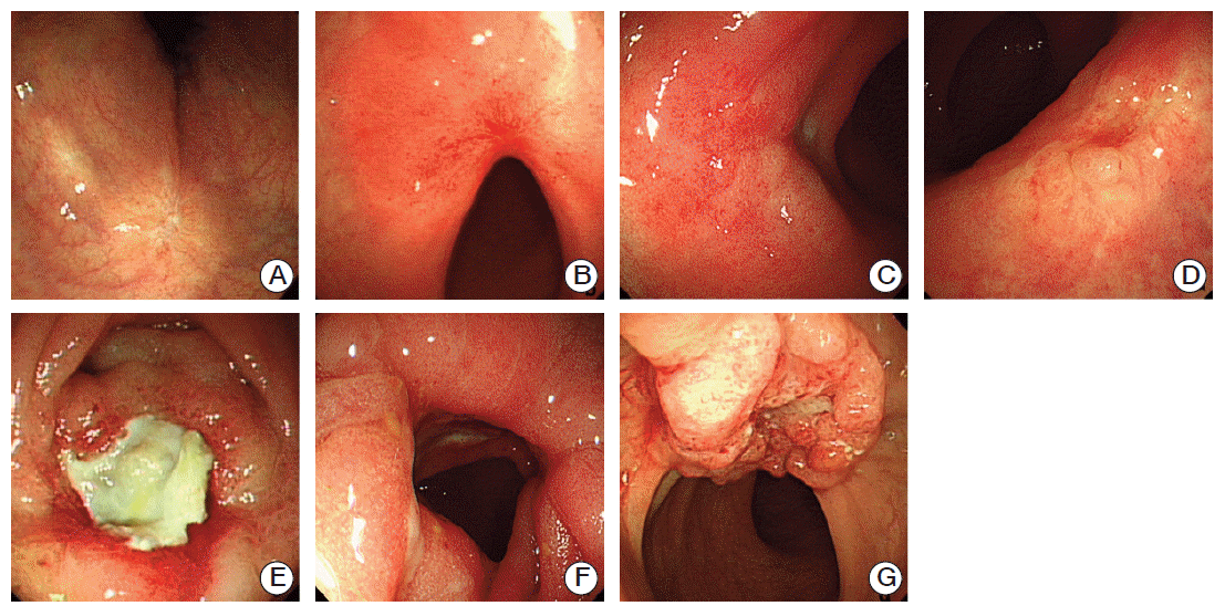
The 481 enrolled patients were randomized into two patient cohorts using a computer generated random sampling, with 193 included in the testing set and 288 included in the validation set. Three expert endoscopists (A, B, and C) evaluated the endoscopic findings based on recorded endoscopic pictures, and determined by consensus the standard endoscopic findings after preoperative CRT based on previous reports [12-14]. These findings included scarring, telangiectasia, erythema, nodules, ulcers, strictures, and remnant tumor, as defined in Table 1.
1. Testing set
Endoscopist A performed evaluation of the testing set. Preoperative endoscopic findings and postoperative histopathologic results of 193 randomly selected patients were reviewed. Following analysis of the endoscopic characteristics used to differentiate between p-GR and p-MR, the patients were classified according to two groups: those with endoscopic findings for p-GR (e-GR) and p-MR (e-MR).
2. Validation set
Endoscopists B and C evaluated the validation set. These two endoscopists reviewed and classified endoscopic findings of 288 patients without information on their ypT classification, using the endoscopic criteria determined in the testing group. Agreement between e-GR and p-GR was analyzed.
3. Statistical analyses
Continuous variables were compared using Student’s t test and categorical variables using the chi-squared or Fisher exact test, as appropriate. The validity of the endoscopic criteria was determined by calculating kappa value, sensitivity, specificity, positive predictive value (PPV), negative predictive value (NPV), and positive and negative likelihood ratios.
p-values of < 0.05 were considered statistically significant. All statistical analyses were performed using SPSS ver. 14.0 (SPSS Inc., Chicago, IL).
Results
The 481 included patients consisted of 344 men and 137 women, with a mean age of 59.7 years (range, 27 to 87 years). Of these, 478 patients underwent radical resection and three patients underwent local excision. Mean tumor sizes were 5.5 cm (range, 1.5 to 12 cm) before CRT and 2.7 cm (range, 0 to 9.5 cm) after CRT. Of these patients, 53 (11.0%) had ypT0 tumors, nine (1.9%) had ypTis, 26 (5.4%) had ypT1, 112(23.3%) had ypT2, 256 (53.2%) had ypT3, and 25 (5.2%) had ypT4 (Table 2).
1. Testing set
Three endoscopic characteristics were associated with p-GR (e-GR): scarring, telangiectasia, and erythema (Table 3, Fig. 1), while four endoscopic characteristics were associated with p-MR (e-MR): nodule, ulcer, stricture, and remnant tumor (Fig. 1). The sensitivity and specificity of these endoscopic characteristics in predicting p-GR were 0.538 and 0.935, respectively, whereas the PPV and NPV were 0.677 and 0.889, respectively.
2. Validation set
Using the endoscopic criteria determined in the testing set, classification of the 288 patients in the validation set was performed by two endoscopists (Table 4). The kappa statistic for inter-observer agreement was 0.965 (p < 0.001), and the sensitivity and specificity of the endoscopic criteria in predicting p-GR were 0.362 and 0.963, respectively, for endoscopist B and 0.277 and 0.959, respectively, for endoscopist C. The PPV and NPV of these criteria were 0.677 and 0.889, respectively, for endoscopist B and 0.654 and 0.885, respectively, for endoscopist C. The false-positive and false-negative ratios were 0.323 and 0.111, respectively, for endoscopist B and 0.346 and 0.115, respectively, for endoscopist C (Table 5).
Discussion
Several recent studies have reported on the feasibility of local excision of selected down-staged rectal cancers after preoperative CRT. In determining whether local excision is feasible in individual patients, proper evaluation of ypT after preoperative CRT is essential. Methods normally used for evaluation of ypT include digital rectal examination (DRE), imaging modalities (e.g., magnetic resonance imaging [MRI], ultrasound), and endoscopy. DRE is a simple and effective method of evaluating distal rectal lesions. Although very effective in detection of primary tumors, it has been found to underestimate the extent of rectal cancer response after preoperative CRT, suggesting that ypT should not be evaluated solely by DRE [15,16]. Ultrasound and MRI are also commonly used for preoperative evaluation of tumor stage in rectal cancer patients; however, evaluation by ultrasound was reported to be less accurate in assessing rectal tumor response after preoperative CRT [17], and MRI could not predict pathological complete response after preoperative CRT [12]. Serum concentration of chorioembryonic antigen (CEA) and re-biopsy may be useful for predicting pathologic CR after preoperative CRT; however, evidence is still lacking [12,13].
Currently, endoscopy is the only method to enable direct evaluation of rectal cancers after CRT, suggesting that it may be more effective for evaluation of ypT than other methods [12,14]. However, despite its efficacy, endoscopic descriptions remain subjective and non-standardized. Thus, there is a need for standardized criteria for endoscopic evaluation of ypT. In this study, the endoscopic findings of post-CRT rectal cancer were categorized according to seven categories—scarring, telangiectasia, erythema, nodule, ulcer, stricture, and remnant tumor—and these categories were used in classifying patients according to two response groups (e-GR and e-MR). Agreement between e-GR and p-GR was also analyzed. The kappa statistic of our endoscopic criteria was excellent (0.965), indicating that these criteria can be easily applied. We also found that this classification system has very high specificity (0.963) and NPV (0.885), but relatively low sensitivity (0.362) and PPV (0.654), suggesting that these endoscopic criteria can strongly predict p-MR cases, but are less able to predict p-GR. Thus, these criteria can be helpful in selection of candidates for local excision.
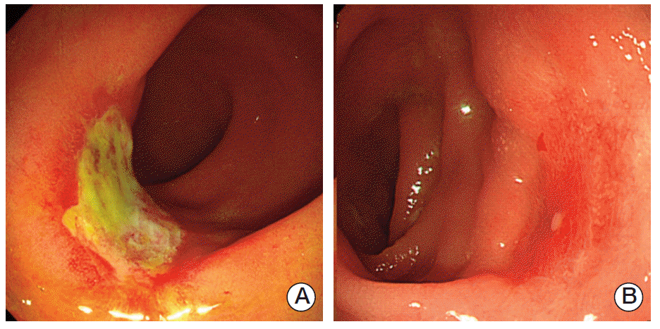
Residual cancer cells in rectal cancer specimens after chemoradiation have been reported to be preferentially located close to the invasive front, not the mucosal surface [18], suggesting that endoscopic findings are not predictive of ypT0. Therefore, our classification system had high specificity, not sensitivity. In addition, some cases showed endoscopic findings not matching the pathologic response (Fig. 2). Development of a more accurate prediction system, with higher sensitivity, requires additional studies that include other methods of evaluation, including MRI, ultrasound, CEA, and/or clinical factors.
This study had several limitations. The study was retro-spective in design and used recorded endoscopic pictures, some of which were limited. In addition, the number of patients who showed clinical or p-GRs may have been insufficient for data analysis. Conduct of large prospective studies will be required in order to validate our novel endoscopic criteria. Another limitation is that lymph node involvement of tumors was not considered in this study. The contraindication for local excision includes rectal cancers with lymph node metastasis. Therefore, evaluation of the lymph node status using imaging modalities is necessary prior to application of these criteria in clinical practice.
Conclusion
In conclusion, this study describes simple and objective endoscopic criteria for evaluation of the ypT of rectal cancer following preoperative CRT. These endoscopic criteria may be narrowly used to determine the suitability of patients for local excision, with or without additional radical surgery, following down-staging after preoperative CRT, since patients who do not show a pathologic good response could be ruled out.